Download
1 / 63
820 likes | 1.35k Views
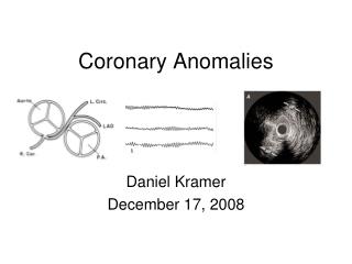
Coronary Artery Anomalies. Dr Rajesh K F. CAAs has a global incidence of 5.64% I ncidence of CAA related sudden death is 0.6%. Normal Features of Coronary Anatomy. CLASSIFICATION OF CORONARY ANOMALIES. Anomalies of origination and course. Absent left main trunk (split origination of LCA )

E N D
Coronary Artery Anomalies Dr Rajesh K F
CAAs has a global incidence of 5.64% Incidence of CAA related sudden death is 0.6%
Anomalies of origination and course Absent left main trunk (split origination of LCA) • LAD and Cx originates from LCS without common trunk • Occurs in about 1% • More frequent with BAV • No clinical consequences • Coronary ostia are smaller • JL catheter for selective cannulation of LAD and amplatz left catheter for LCX
Anomalies of origination and course Anomalous location of coronary ostium within aortic root or near proper aortic sinus of Valsalva a. High b. Low c. Commissural • High origin of LCA engaged with Amplatzleft,MP catheter • High origin of RCA engaged with Amplatz catheter
Anomalies of origination and course Anomalous location of coronary ostium outside normal aortic sinuses a. Right posterior aortic sinus b. LV ,RV ,PA ,AA ,Aortic arch, Descending thoracic aorta, Innominate artery ,Right carotid artery ,IMA , Bronchial artery ,Subclavian artery c.Pulmonary artery
Ectopic Coronary Ostium in NCS • Benign anomaly • Most commonly involves LCA ostium • Difficult cannulation-unexpected location,tangential or slitlike nature • Nonselective angiography-longer than usual LMCA • RAO and straight lateral projections are most useful • For selective catheterization Amplatz or Multipurpose curved catheters offer best chance of success
Ectopic Coronary Ostium Arising Outside the Aortic Root(Ascending Aorta) • Usually involves anterior/left surface of aorta • Frequently have slit like orifices and tangential proximal course along aortic wall • RCA is most frequently ectopic artery • Predisposed to more atherosclerosis • Association with Congenital aortic valve anomalies
Ectopic Coronary from Pulmonary artery (1) LCA from posterior facing sinus (2) Cx from posterior facing sinus (3) LAD from posterior facing sinus (4) RCA from anterior right facing sinus (5) Ectopic location (outside facing sinuses) (a) From anterior left sinus (b) From pulmonary trunk (c) From pulmonary branch Smith A., Arnold R., Anderson R.H., Wilkinson J.L., Qureshi S.A., McKay R. Anomalous origin of the left coronary artery from the pulmonary trunk. J ThoracCardiovascSurg 1989;98
ALCAPA • LCA arises from PA usually from left posterior facing sinus • Fetus-both coronary arteries receive forward flow • Early after birth - Anterolateral infarct and slight retrograde flow from LCA to PA • 15% of patients-myocardial blood flow can sustain myocardial function at rest or even during exercise • Adult-Enlarged RCA and collaterals and significant retrograde flow into PA
Clinical features Paroxysmal attacks of acute discomfort precipitated by feeding CHF at 2 to 3 month Physical examination-CHF,MR Abnormal Q waves in leads I, aVL, and precordial leads V4 to V6 Older children and adults -may be asymptomatic or have dyspnea, syncope, or exertional angina Sudden death after exertion
Echo • Abnormal origin of LCA • Flow passes from RCA into PA • Enlarged RCA • RWMA and mitral regurgitation Aortic root angiography • Dilated RCA • large collaterals-filling of LCA and passage of contrast material to MPA
Treatment • Ligation of LCA at its origin • Direct reimplantation of origin of LCA into aorta (with a button of PA around the origin) • Ligation of origin of LCA and reconstitution of flow through it with subclavian arterial or SVG • Takeuchi procedure- aortopulmonary window is created and a tunnel fashioned that directs blood from aorta to LCA
Anomalous Origination of RCA, LAD or Cx Artery From PA • Benign condition • Typically recognized by atypical angina, systolic heart murmur , abnormal stress test or angiography • In absence of major clinical manifestations not an indication for surgery
Anomalies of origination and course Coronary ostium at improper sinus (ACAOS) a. RCA from left anterior sinus b. LAD from right anterior sinus c. Cx from right anterior sinus d. LCA from right anterior sinus ARCA~ 6 times more prevalent than ALCA ALCA has a higher risk of SCD
Anomalies of origination and course Sudden cardiac death associated with four risk factors • Slit-like coronary orifice • Acute angle of take-off from aorta • Presence of aortic intramural coronary arteries • Inter-arterial course between aorta and PA
Echo • Difficult by angiography • MRI or CT is more sensitive
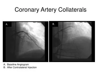
Abnormal crossing pathways • Retrocardiac • Path is behind mitral and tricuspid valves in posterior AV groove Retrocardiac Angelini et al
Retroaortic • Most common • Specifically involve origination of Cx artery from right sinus of Valsalva • Incidence in general population range from 0.1 to 0.9% • No clinical consequences
Preaortic • Between the aorta and PA • Osial abnormalities are usual • intramural course within the aortic root and adherent to it for about 1.5 cm • Rarely systolic compression lead to SCD
Intraseptal(supracristal) • Located in upper anterior IVS(Derived from conal septum) • Recogonised by systolic phasic narrowing during CAG • Septalperforators
Prepulmonary (precardiac) • Common in TOF
RCA from left anterior sinus, with anomalous course(ARCA) (1) Posterior AV groove or retrocardiac (2) Retroaortic (3) Between aorta PA (Most common, 30% mortality) (4) Intraseptal (5) Anterior to pulmonary outflow (6) Posteroanteriorinterventricular groove (wraparound)
LCA from right anterior sinus, with anomalous course (1) Between aorta and PA (2) Anterior to PA (3) Retroaortic (4) Intraseptal (5) Posterior AV groove (6) Postero-anterior interventricular groove
LAD from right anterior sinus, with anomalous course (1) Anterior to PA (2) Intraseptal (3) Between aorta and PA (4) Posteroanteriorinterventricular groove (wraparound)
Cx that arises from right anterior sinus, with anomalous course (1) Posterior AV groove (2) Retroaortic Angiography • Long LMCA segment • Small cx branch • RAO view shows dot of retro aortic course
Current recommendations Symptomatic patients • Unroofing • Reimplantation Asymptomatic patient • Demonstrable ischemia: repair • No demonstrable ischemia • ALCA: repair • ARCA: observe
Anomalies of origination and course Single coronary artery • Incidence 0.024% • Single ostium with absence of an ostium in opposite sinus • No other coronary artery from an ectopic site • 40% associated with cardiac malformations(TGA, TOF, TA, coronary-cameral fistulas, and BAV)
Less tangential origin or ostial ridge pathology than ectopic CA with independent ostia • Coronary blood flow is not affected • Incidence of atherosclerotic disease not increased • Ostialobstruction (large guiding catheter or directional atherectomydevice) -poorly tolerated • Angioplasty of common trunk is absolutely contraindicated
Angiographic classification Lipton et al
Coronary artery atresia • Characterized by ostialdimple in left or right aortic sinus terminates in a cordlike fibrotic structure without a patent lumen • Proximal occluded segments have larger diameter • Collaterals • Stress testing demonstrate myocardial ischemia • Seen in PA with intact IVS
Coronary artery isolation Variant of ostial atresia Juxtaposition of abnormal aortic cusp with aortic sinus wall lead to obliteration of underlying coronary ostium
Anomalies of intrinsic anatomy Coronary hypoplasia Angiographic appearance -small diameter(usually <1 mm) with respect to apparent area of dependent myocardium Myocardial fibrosis and segmental hypokinasia are frequent Local reversible ischemia during stress testing with myocardial nuclear scintigraphy Terminology is incorrect when dependent myocardial bed is actually served by alternative sources (unusual coronary patterns) or coronary spasm or diffuse disease
Anomalies of intrinsic anatomy Absent coronary artery Misnomer On angiographic grounds, the most frequent reason are Coronary ectopia (misdiagnosed) Coronary occlusion with lack of demonstrable collateral filling Alternative coronary artery tree pattern not recognized on angiography
Anomalies of intrinsic anatomy Coronary ectasia or aneurysm Localized dilations in a normal sized coronary artery Coronary segment diameter >50% with respect to normal Doppler coronary wire blood flow velocity - significant reduction in peak flow velocity Streamlining and slow runoff contrast media in angiography Acquired stenosis Mural thrombosis with distal embolization Aneurysmal rupture
Intramural coronary artery (muscular bridge) • More than 1% of incidence • Most commonly associated with ventricular hypertrophy • Coronary artery segment of variable length covered by myocardial fibers • Angiographic recognition of systolic narrowing • Phasic narrowing of a coronary aftery may also occur in ventricular aneurysms or pericardial fibrous bands • Intracoronary nitroglycerin in a 100- to 300-/microg bolus facilitates
U sign- Artery's accentuated descent from its subepicaardial location • Most commonly involve proximal LAD • Systolic stenosis is unlikely to cause coronary flow reduction • Rare reports of spasm, thrombus and atherosclerotic change
Anomalies of intrinsic anatomy • Subendocardialcoronary course • Coronary crossing • Anomalous origination of posterior descending artery from anterior descending branch or a septal penetrating branch
Anomalies of intrinsic anatomy Split RCA a. Proximal+distal PDAs that both arise from RCA b. Proximal PDA that arises from RCA, distal PDA arises from LAD c. Parallel PDAs (arising from RCA, Cx) or “codominant”
Anomalies of intrinsic anatomy Split LAD Duplication of LAD < 1 % Population a. LAD+first large septal branch b. LAD, double (parallel LADs) c. LAD+large diagonal d. Highly dominant RCA+LAD
Anomalies of intrinsic anatomy Ectopic origination of first septal branch a. RCA b. Right sinus c. Diagonal d. Ramus e. Cx
Anomalies of coronary termination Coronary fistulas • A sizable communication between a coronary artery and a cardiac cavity or any segment of systemic or pulmonary circulation
Anomalies of coronary termination Fistulas from RCA, LCA, or infundibular artery to RV,RA,CS , SVC, PA, PV,LA, LV, Multiple(right+left ventricles) Originate from left coronary artery system (50-60%), right coronary artery system (30-40%), or both (2-5%) Most fistulas (90%) drain into right heart
Functional coronary fistula • Definite signs of fistulous flow-feeding vessel diameter >50% expected diameter • Angiographically prompt visualization of receiving structure with step up in concentration of injected substance • Evidence of volume overload in affected cardiac chambers • Evidence of steal involving myocardial nutrient blood flow
Use large-lumened catheters with side holes (NIH or Gensini ) or guide catheter kept in position by a 0.014-inch guidewire Mechanical injector Diagnose coronary fistula Identify receiving chamber or vessel Complete visualization of nutrient myocardial branches Absence of nutrient coronary branches arising from a fistulous tract should suggest alternative diagnosis-RSOV
Complications Aneurysm formation Intimal ulceration Medial degeneration Intimal rupture Atherosclerotic deposition Calcification Side branch (nutrient) obstruction Mural thrombosis Coronary rupture