Download
1 / 54
540 likes | 775 Views
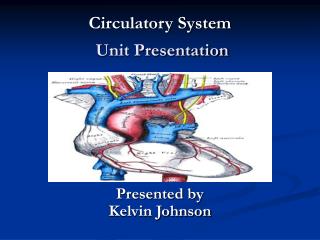
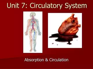
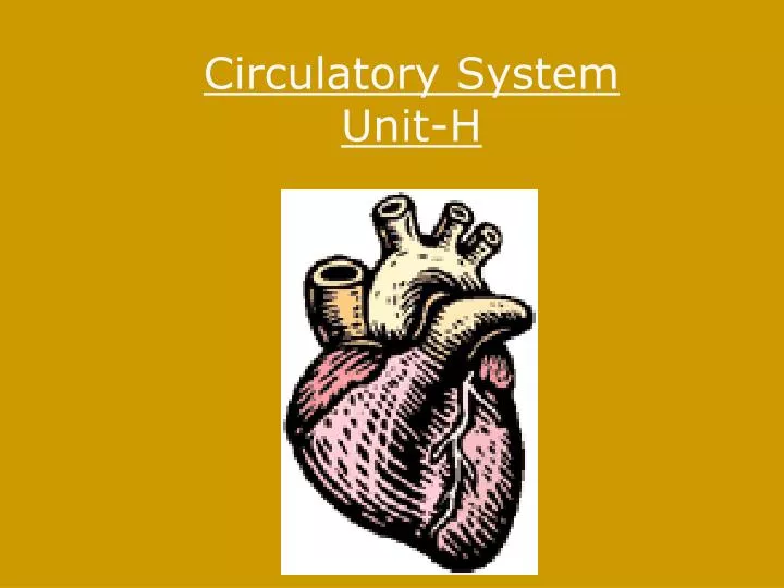
Circulatory System Unit-H. Functions Pump Blood transport system around the body Carries O2 and nutrients to cells, carries away waste products Lymph system Lymph system – returns excess tissue fluid to general circulation. Circulatory System. Structures and Circuits. Structures

E N D
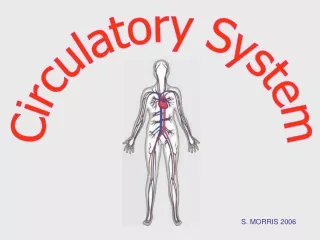
Functions Pump Blood transport system around the body Carries O2 and nutrients to cells, carries away waste products Lymph system Lymph system – returns excess tissue fluid to general circulation Circulatory System
Structures and Circuits Structures • Heart, Arteries, Veins, Capillaries. • Blood and lymph are part of circulatory system. Major Blood Circuits • Cardiopulmonary Circulation-Circulationof blood to the lungs to pick up O2. • Systemic Circulation- from the heart to the tissues and cells, then back to the heart
Circulation from the heart to the tissues and cells, then back to the heart. Systemic Circulation
Circulationof blood to the lungs to pick up O2. Cardiopulmonary Circulation
Goal of the Cardiovascular System: deliver blood to all parts of the body • Does so by using different types of tubing, attached to a pulsatile pump • Elastic arteries • Muscular arteries • Arterioles • Capillaries • Venuoles • Veins • Distribution system broken up into areas called vascular beds • Skin • Digestive (splanchnic) • Muscle
The Heart • Muscular Organ • Size of a closed fist • Weighs 12-13 oz • Location – thoracic cavity • Apex- conical tip, lies on diaphragm, points left. • Stethoscope- instrument used to hear the heartbeat.
Hollow, muscular, double pump that circulates blood. At rest = 2 oz with each beat, 5 quarts./min., 75 gallons per hour. Ave = 72-75 beats per minute 100,000 beats per day PERICARDIUM- double layer of fibrous tissue that surrounds the heart. MYOCARDIUM- cardiac muscle tissue ENDOCARDIUM- smooth inner lining of heart SEPTUM- partition (wall) that separates right half and left half. Heart Structure
Superior Vena Cava and Inferior Vena Cava- bring deoxygenated blood to right atrium. Pulmonary artery- takes blood away from right ventricle to the lungs for O2 Pulmonary veins- bring oxygenated blood from lungs to left atrium. Aorta- takes blood away from left ventricles to rest of the body.
Chambers and Valves Septum divides into Rt. & Lt. halves. Upper chambers- Rt. & Lt. ATRIUMS Lower chambers- Rt. & Lt. VENTRICLES Four heart valves permit flow of blood in one direction. .
TRICUSPID VALVE- between right atrium and right ventricle. BICUSPID (MITRAL) VALVE-between left atrium and left ventricle Semi lunar valves are located where blood leaves the heart – PULMONARY SEMILUNAR VALVE AORTIC SEMILUNAR VALVE- Prevents backflow of blood as it leaves the heart. Artia- Upper chambers of the heart.
Physiology of the Heart The Heart is a double pump. When the heart beats…. Right Heart Deoxygenated blood flows into heart from vana cava > right atrium > tricuspid valve > right ventricle > pulmonary semilunar valve > pulmonary artery > lungs (for oxygen) Left Heart Oxygenated blood flows from lungs via pulmonary veins > left atrium > mitral valve > left ventricle > aortic semilunar valve > aorta > general circulation (to deliver oxygen) • Heart Sounds = lubb dupp
SA (sinoatrial) NODE = PACEMAKER Located in right atrium SA node sends out electrical impulse Impulse spreads over atria, making them contract Travels to AV Node AV (atrioventricular) NODE Conducting cell group between atria and ventricle Carries impulse to bundle of His BUNDLE OF HIS Conducting fibers in septum Divides into R and L branches to network of branches in ventricles (Purkinje fibers) PURKINJE FIBERS Impulse shoots along Purkinje fibers causing ventricles to contract Control of Heart Contractions
Device used to record the electrical activity of the heart. ELECTROCARDIOGRAM (EKG or ECG)
SYSTOLE = contraction phase DIASTOLE = relaxation phase P = atrial contraction QRS = ventricular contract T = ventricular relaxation
Circulation and Blood Vessels ARTERIOLES- small arteries (highest level of 02) VENULES- small veins
AORTA- largest artery in the body • First branch is coronary artery • Aortic arch • Many arteries branch off the descending aorta
Carry deoxygenated blood away from capillaries to the heart Veins contain a muscular layer, but less elastic and muscular than arteries Thin walled veins collapse easily when not filled with blood VEINS
VALVES- permit flow of blood only in one direction of the heart. JUGULAR vein- located in the neck
Smallest blood vessels, can only be seen with a microscope Connect arterioles with venules Walls are one-cell thick and extremely thin- allow for selective permeability of nutrients, oxygen, CO2 and metabolic wastes. Made up of Endothelial Cells. CAPILLARIES
Carry oxygenated blood away from the heart to the capillaries Elastic, muscular and thick-walled Transport blood under very high pressure The major artery that carries blood to the brain is the Carotid. ARTERIES
Surge of blood when heart pumps creates pressure against the walls of the arteries. SYSTOLIC PRESSURE- measured during the contraction phase. DIASTOLIC PRESSURE- measured when the ventricles are relaxed. Average systolic = 120 Average diastolic = 80 PULSE- alternating expansion and contraction of an artery as blood flows through it. Blood Pressure http://www.phschool.com/science/biology_place/labbench/lab10/images/bloodpr.gif
Diseases of the Heart • ARRHYTHMIA (or dysrrhythmia) - any change from normal heart rate or rhythm.
BRADYCARDIA – slow heart rate (<60 bpm) • TACHYCARDIA – rapid heart rate (>100 bpm)
Coronary Artery Disease ANGINA PECTORIS- chest pain, caused by lack of oxygen to heart muscle, treat with nitroglycerin to dilate coronary arteries.
MI or heart attack Lack of blood supply to myocardium causes damage Due to blockage of coronary artery or blood clot atherosclerosis- plaque build up on arterial walls, or arteriosclerosis- loss of elasticity and thickening of wall. Amount of damaged depends on size of area deprived of oxygen. Symptoms – severe chest pain radiating to left shoulder, arm, neck and jaw. Also nausea, diaphoresis, dyspnea. Immediate medical care is critical Rx- bed rest, oxygen, medication Morphine for pain, TPA to dissolve clot MYOCADIAL INFARCTION
Morphine for pain, Tissue Plasminogen Activator (tPA)to dissolve clot
Anticoagulant therapy to prevent further clots from forming. • Angioplasty and by-pass surgery may be necessary
Heart Surgery • CORONARY BY-PASS – usually, a healthy vein from the leg or arm is removed and attached before and after the coronary obstruction, creating an alternate route for blood supply to the myocardium
PACEMAKERS Demand pacemaker- fires only when heart rate drops below programmed minimum.
AED AED is a portable electronic device that automatically diagnoses the potentially life threatening cardiac arrhythmias of ventricular fibrillation and ventricular tachycardia. The application of electrical therapy which stops the arrhythmia, allowing the heart to re-establish an effective rhythm.
Electrical shock to bring the heart back to normal rhythm. DEFIBRILLATION
CPR • Cardiopulmonary resuscitation, used in the presence of cardiac arrest.
Disorders of the Blood Vessels • Aneurysm- ballooning of an artery, thinning and weakening.
Swollen, distended veins- heredity or does to posture, prolonged periods of standing, physical exertion, age and pregnancy Varicose Veins
Disorders Cont. • HYPERTENSION • High blood pressure • “silent killer” – usually no symptoms • Condition leads to strokes, heart attacks, and kidney failure • 140/90 or higher • Higher in African-Americans and post-menopausal women • Risk factors = smoking, overweight, stress, high fat diets, family history • Treatment = Relaxtion, low fat diet, exercise, weight loss, medication HYPOTENSION- Low blood pressure, systolic<100