Download

1 / 8
80 likes | 147 Views
This informative guide outlines the essential structures of fetal circulation such as Ductus Arteriosus, Foramen Ovale, Ductus Venosus, Umbilical Artery, and Umbilical Vein. Learn the critical variances and functions of these key components in the fetal circulatory system.

E N D
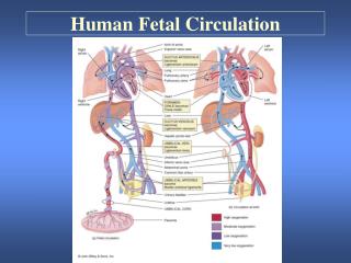
Fetal Circulation Some key features & differences…
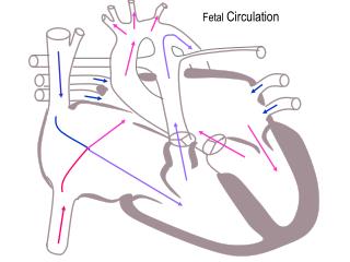
Important Structures • DuctusArteriosus • Runs from pulmonary artery to aorta • Bypasses lungs in fetus • Collapses at birth due to demand of lungs for blood
Foramen Ovale • Left & right atria both have a “hole” in inside wall • Allows blood to flow from right to left atria bypasses lungs! • Expansion of left atrium at birth puts 2 holes out of alignment
DuctusVenosus • Stops carrying blood from moment of birth • Channel through liver that carries blood from placenta to inferior vena cava • Approx ½ blood enters liver • Approx ½ blood bypasses liver ductus venosus inferior vena cava heart
Umbilical Artery • Carries blood LOW in oxygen/nutrients from baby to mom • Umbilical Vein - Carries blood RICH in oxygen/nutrients from mom to baby
Key Differences • Umbilical vein transports oxygen rich blood to the fetus • Fetal blood has a much greater capacity to carry oxygen • Vein carries blood to the liver and is partially shunted by the ductusvenosus this allows about half the blood to bypass the liver! • The ductus joins the inferior vena cava • Blood from the lower part of the fetal body is mixed with oxygen rich blood from the placenta
Blood is forced into the right atrium. • The fetal heart has an opening between the right and left atria that shunts blood away from the lungs. • This opening is called the foramen ovale. • The ductusarteriosus conducts blood away from the pulmonary trunk to the aorta (lung bypass).