Download
1 / 29
300 likes | 365 Views
NO Detection via Chemiluminescence and Fluorescence Martin Feelisch, Ph.D. Boston University School of Medicine Department of Medicine/Section of Molecular Medicine Whitaker Cardiovascular Institute. “Rigorous Detection and Identification of Free Radicals In Biology and Medicine”, Workshop,

E N D
NO Detection via Chemiluminescence and Fluorescence Martin Feelisch, Ph.D. Boston University School of Medicine Department of Medicine/Section of Molecular Medicine Whitaker Cardiovascular Institute “Rigorous Detection and Identification of Free Radicals In Biology and Medicine”, Workshop, 12th Annual Meeting of the Society for Free Radical Biology and Medicine, Hilton, Austin, Nov 16, 2005
Part I. NO Detection Using Gas Phase Chemiluminescence
Advantages and Disadvantages of Chemiluminescence Compared to Other Methods of NO Detection · Advantages High Sensitivity and Linearity over a Broad Concentration Range Good Reproducibility and High Specificity for NO(few other gaseous substances (DMSO, ethylene) react with ozone) Measurements Possible in Turbid or Colored Samples, Even at Extreme pH(in solution, in the headspace, in expired air) Besides Mass Spectrometry the Only Other Method that Allows Quantification of Absolute Amounts of Nitroso Species Moderate Running Costs Disadvantages With Some Biological Samples Difficult to Extract NO into Gas Phase Provides Limited Structural Information Limited Sample Throughput, High Purchase Price for Detector ·
Quantification of NO, Nitroso and Nitrosyl Species Using Gas Phase Chemiluminescence NO present or generated in an aqueous system has to be purged out of solution by an inert gas (N2, Ar, He) to be available for analysis in the gas phase. Biological samples containing nitroso and nitrosyl compounds are processed using either Photolysis to cleave NO-adducts or Chemical Reaction (i.e., injection into a denitrosating reaction mixture) to convert these species into NO. The NO-containing gas is then transfered to the analyzer for quantification. Detection Principle: In the reaction chamber NO is mixed, at a defined flow rate and under reduced pressure, with ozone. NO + O3 NO2* + O2 NO2* NO2 + h . The light that is emitted by the fraction of excited NO2 on returning to the ground state (chemiluminescence; 640-3000 nm) is measured by photon counting. With O3 being present in large excess, light intensity is directly proportional to [NO]
Photolysis/Chemiluminescence Approach for Detection of Nitroso Species Reaction principle: RSNO RS. + NO. R2NNO R2N.+ NO. Discrimination of RSNOs from other nitroso species and nitrite by measurement before and after HgCl2 treatment, and with lamp ON and OFF Stamler et al., 1992, Alpert et al., 1997 light light
Advantages and Problems with Photolysis-Based Techniques Advantages Low interference by contamination with nitrite Fewer interferences with components of the redox-active reaction mix Problems Extremely high temperatures are reached inside of the photolysis cell Reported RSNO values using photolysis-based technques are orders of magnitudehigher than those using reductive methods(µM rather than nM) Artefactual generation of nitroso species by the photolysis of nitrate (NO3-) and the trapping of RNOS by thiols (Dejam et al., FRBM, 2004) Photolysis of nitroso species other than S-nitrosothiols also generates a signal Controls with nitrite and mercuric chloride are pointless due to low photolysis yield of nitrite and the ability of mercuric salts to complex sulfhydryl groups ( blockage of the targets of artefactual nitrosation naturally leads to lower levels of nitroso-related signals in the presence of HgCl2, but this does not neccessarily indicate involvement of RSNOs)
Sample Processing Using Redox-Active Reaction Mixtures Many Choices, Many Pitfalls Most techniques use Chemical Reactions to convert nitroso and nitrosyl species into NO, which is then detected by chemiluminescence Reducing mixturesdiffer largely in reducing strengths and reduction capacity Iodine/iodide (I3-) 60 mM I-/6-20 mM I2/ 1M HCl, RT Samouilov & Zweier, 1998 56 mM I-/ 2 mM I2, 4mM CuCl, CH3COOH, 68°C Marley et al., 2000 60 mM I-/10 mM I2, CH3COOH, 60°C Feelisch et al., 2002 Cysteine/CuCl 1 mM L-cysteine, 0.1 mM CuCl Fang et al., 1998 Hydroqinone/Quinone 0.1/0.01 mM Samouilov & Zweier, 1998 VCl3/H+ 0.1 M in 2M HCl Ewing et al., 1998 Oxidizing mixture for determination of NO-hemes Ferricyanide 0.2 M in PBS pH 7.5 Gladwin et al., 2002 Bryan et al, 2004 General Problem: Neither method is absolutely specific and bears the potential to produce false positive (nitrate, L-NitroArg, ...) or negative signals
Reaction Chambers Come in Many Different Designs Menon et al., 1991 Cox & Frank, 1982 Dunham et al., 1995
The Most Frequently Used Type of Chemiluminescence Set-up Samouilov & Zweier, 1998
Application Example Direct Measurement of NO Release from NO Donor Compounds expected: 100 pmol found: 107 pmol 1 µM MAHMA/NONOate (direct injection of 100 µL into PBS, 37°C)
Which NO-Related Species are Detected and How can they be Discriminated from One Another? Without Reduction Step: NO (direct injection into buffer or water) Upon Acidification: NO2-(disproportionation of HNO2) RONO (acid-catalysed decompos.) With Sample Reduction: NO2-/NO3-(KI/CH3COOH, RT for nitrite, VCl3/H+, 90°C for nitrate) RSNO RNNO(I3-/CH3COOH, 60°C) NO-Heme detection limit: 1-50 nM, depending on flow and inj. volume 250 fmoles NO (@ 50 µL injection vol.) Discrimination between different species: Selective NO2-removal Sulfanilamide/H+ RSNOs from other Nitroso-Species HgCl2/sulfanilamide Nitroso from Nitrosyl Species Reducing vs. Oxidizing Reaction Mix
Now, how am I going to do this practically? biological sample Split into aliquots Nitrosyl Species Nitrite and Nitroso Species (direct injection) (split into further aliquots, depending on requirements, differentiation between different types of compounds by reaction with group-specific reagents) Oxidation Reduction (Ferricyanide Solution, (Iodine/Iodide Reaction Mix, neutral pH) acidic pH)
Application Example Detection of Nitrosyl-Heme Species in Rat Tissues Using Ferricyanide Brain Ferricyanide Fe3+ Fe2+ Fe3+ NO-Hb MetHb + NO Ferrocyanide Fe2+ RBC lysate Plasma Heart Liver Kidney Lung Aorta 5 10 15 20 25 30 35 40 0.05M ferricyanide in PBS, pH 7.5 @ 37°C
1 2 3 untreated sample sulfanilamide/H+ (15 min) HgCl2+sulfanilamide/H+ (45 min) nitrite RSNO other nitroso compounds nitrite RSNO other nitroso compounds nitrite RSNO other nitroso compounds Discrimination Between Nitrite and Different Nitroso Species Using a Reducing Iodine/Iodide Reaction Mix in Combination with Group-Specific Reagents biological sample
Discrimination Between Nitrite andNitroso Species Using Group-Specific Sample Processing 1 2 3 + + HgCl2/ Sulf/H+ 30 untreated +Sulf/H 20 Nitrite NO [ppb] 10 RSNO RNNO (and possibly other species) 0 0 5 10 15 Time [min]
Thiol Alkylation and Nitrite Removal are Required to Prevent Artifactual Nitrosation of Cellular Constituents Problem: Reaction of protein thiols (or GSH) with nitrite to form RSNO Solution: Alkylation of –SH groups with either NEM or iodacetamide (5-10 mM in PBS, 15 min) Nitrite Removal using Sulfanilamide/H+ (15 min RT; azide, urea, sulfamic acid don’t work!) using Size Exclusion Chromatography (Sephadex G-25), followed by Sulfanilamide/H+ SNO SNO + NO2- HS HS SH SNO
Most Important Factors Affecting Assay Sensitivity Injection Volume 10-1000 µL, depending on sample availability, size of reaction vessel and gas flow Rate of Reduction Molarity and Temperature of the Reaction Mixture (rapid reduction produces sharp peaks) Flow Rate of Purging Gas and Dead Space of the System 50 mL/min-3000 mL/min (depending on whether developed for environmental monitoring or research) 100-200 µL/min represents a good compromise between short analysis time (high flow) and high sensitivity (low flow) Dead space should be as small as possible Detector Sensitivity, Integration Time and Baseline Noise Largely determined by instrument noise (dependent on photomultiplier type and temperature as well as on reaction chamber design; Dasibi, Sievers and EcoPhysics machines differ by a facor of 2-10) Longer integration time increases sensitivity Baseline noise increases with fluctuations in pressure Nitrite Background Nitrite contamination (water, glass- and plasticware incl. pipette tips, ultrafiltration membranes)
Recent Challenges to the Validity of This Analytical Method Mercury-stable nitroso signal in the iodine/iodide assay may be nitrated lipids rather than N-nitrosamines (Schopfer et al., J Biol Chem 2005) However, spiking with a final concentration of 75 µM (!) of a nitrated lipid standard was required to produce a response similar to the Hg-stable signal in human plasma, while the endogenous concentration of these species was estimated (by the same authors) not to exceed 1-2 µM. The „harsh chemistry“ required to completely remove nitrite from biological samples when working with reductive chemiluminescence-based assay (i.e. the pretreatment with sulfanilamide/H+) renders S-nitrosothiols unstable (Stamler et al., repeated editorial claims without data) (Rogers et al., J Biol Chem, 2005) However, all tested S-nitrosothiols are rock-stable under these conditions (Feelisch et al. & Gladwin et al., unpublished) Heme autocapture may be responsible for the previous lack of detection of nitroso species in human red blood cells (Rogers et al., J Biol Chem, 2005) However, no problem other than peak broadening was observed in the presence of very high conc of Hb (Feelisch et al. & Gladwin et al., unpublished)
Part II. NO Detection & Bioimaging Using Fluorescence
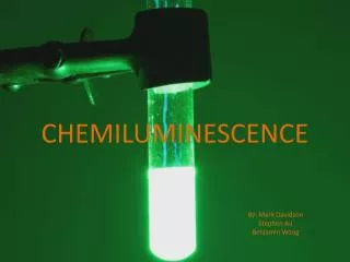
Bioimaging of Nitric Oxide Using DAF-2 Detection principle: Reaction of aromatic vicinal diamines with NO in the presence of oxygen to produce the corresponding triazenes cell membrane DAF-2 DA DAF-2 DAF-2 T NOx esterases fluorescent, Ex 495 nm, Em 515 nm non-fluorescent, cell-permeable non-fluorescent Advantages:Sensitivity for NO (5 nM in vitro) with high temporal and spatial resolution No cross-reactivity to NO2-/NO3- and ONOO- Assay limitations:Possible interference by reducing agents and divalent cations, pH sensitive, subject to photobleaching, requiring standardized illumination conditions
Yes, You can produce lots of pretty images, …
Bioimaging of Nitric Oxide Using DAF-2 in Endothelial Cells Control Unloaded cells Time course of NO formation in response to BK (100 nM) t=0 5s 60s 120s 180s HUVECs P4, labelling: DAF-2 DA 10 µM for 60 min; incubation:HBSS + L-Arg (1 mM), 37°C
Propagation of NO Wave during Stimulation of Endothelial Cells with the Calcium Ionophor, A23187 (1 µM) t=0 0.5 min 1 min 1.5 min 2 min 5 min 10 min after stimulation of cells with BK
Yes, You can produce lots of pretty images, … But …
Problems and Pitfalls with DAF-2 as an NO Probe · Unclear what species exactly is detected in biological systems More likely an indicator of nitrosative events (i.e. of RNOS) than of NO per se The true sensitivity for “NO” in tissues is compromised by the presence of thiols and other antioxidants and autofluorescence of the probe(Rodriguez et al, 2005) Specificity for “subcellular formation of NO” depends on the degree of compartment- alization in the tissue Complex metabolism and susceptibility to oxidation renders quantitative comparisons problematic DAF-2 may undergo oxidative transformation to a radical intermediate(Jourd’heuil; 2002) DAF-2T may undergo rapid reduction or quenching(producing transient signals) DAF-2 forms adducts with ascorbate and dehydroascorbate(Zhang et al, 2002) There is a light-sensitive component in cells/tissues the nature of which is unclear Nitrate/thiol interaction? Formation of adducts with mercuric salts and glutathione results in spectral changes that may be misinterpreted as NO signals (Rodriguez et al, 2005) · · · · · ·
Why does a probe that requires nitrosation work at all in vascular tissue and other biological environments? How does it compete with endogenous antioxidants? How does it compete with other cellular targets (e.g. reactive protein moieties)? Using incubation conditions frequently used in the literature (10 µM) intracellular DAF-2 concentrations approach the millimolar concentration range (Rodriguez et al, 2005)
Compartmentalization of the Probe Around Elastic Lamina Limits its Potential to Characterize the Subcellular Site of NO Production in the Vasculature H&E stain DAF-2 + NO Donor Basal UV light UV illumination leads to levels of nitrosating species that interfere with NO detection by enzymatic sources (Rodriguez et al, 2005)
Can Targets of NO Be Detected Through Photolysis? NO is generated via photolysis from a UV-absorbing species with an absorption peak below 310 nm, consistent with the characteristics of nitrate (NO3-) (Rodriguez et al, 2005)
Recent Developments Development and commercial availability of red fluorescent chromophores (diamino-rhodamine-based; DAR-4M) increases flexibility for combinations with other green-fluorescent probes and shows reduced interference with tissue autofluorescence, but is otherwise very similar to DAF-2 Difluoroboradiaza-s-indacene based fluorophore (similar chemistry) Detection of nitroso peptides and proteins on diaminofluoresceine gels (standard SDS-PAGE followed by UV photolysis in the presence of DAF-2 or DAF-FM for detection of C-, O-, N- ans S-nitrosated compounds) (Mannick et al, 2005) Near-Infrared fluorescent probes for „NO“ detection in isolated organs (tricarbocyanine as NIR fluorochrome coupled to o-phenylenediamine as NO sensor; NIR is potentially very interesting for in vivo imaging approaches as it allows deeper penetration of light into tissues and shows no interference with tissue autofluorescence; promising novel approach (Nagano et al, 2005) Amplifier-coupled fluorescent NO indicator with nanomolar sensitivity in living cells (genetically encoded fluorescent indicator based on the binding of NO to soluble guanyly cyclyase and detection of formed cGMP by FRET; interesting, but potentially problematic cross-talk with cGMP generated by particulate GC and modulation of sensitivity by PDE activity) (Sato et al, 2005)