Download
1 / 42
500 likes | 884 Views
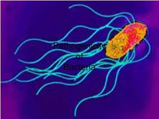
Mechanism of Pathogenicity. Host vs Parasite: Advantage Parasite. Pathogenicity and Virulence. Pathogenicity - ability of MO to cause disease Virulence - degree of pathogenicity; disease-evoking power of MO Measurement - MO’s virulence tested experimentally in animals or in lab

E N D
Mechanism of Pathogenicity Host vs Parasite: Advantage Parasite
Pathogenicity and Virulence • Pathogenicity - ability of MO to cause disease • Virulence - degree of pathogenicity; disease-evoking power of MO • Measurement - MO’s virulence tested experimentally in animals or in lab • LD50 (Lethal Dose) - number MO (or amount toxin) needed kill 50% inoculated hosts (test population) • ID50 (Infectious Dose) - number MO needed to cause disease in 50% test population
Infection, Virulence, Disease • Lower the LD50 or ID50, the more virulent the MO • Likelihood disease results from infection: • Increasing numbers of MO • Decreasing resistance of host
Host Disease Factors • Susceptible (overall health) • Gender (female, male) • Nutritional status (balanced, diet) • Weather and climate (seasons, hot, cold, moisture) • Fatigue (lack of rest, sleep) • Age (very young, old) • Habits (active/inactive, over/under weight) • Life style (physical, mental, social, spiritual) • Pre-existing illness (inherited, chronic, infection) • Emotional disturbance (stress, anger) • Chemotherapy (legal, illegal drugs)
Disease By MO • Must gain entrance to host • Portal of entry - avenue by which MO enters host • Include: • Mucous membrane • Skin • Parenteral route
Entry Mucous Membrane: RT, GI Tract • Respiratory Tract – easiest, most frequent; via aerosols, direct mucous membrane contact (i.e., influenza, pneumonia, TB, measles, smallpox) • Gastrointestinal Tract – ingested via food, water, dirty hands; entry fecal-oral route: • Survive HCl (stomach), bile and digestive enzymes (small intestine) • Exit in feces (i.e. polio, infectious hepatitis, typhoid fever, bacillary dysentery, amoebic dysentery, cholera)
Entry Mucous Membrane: GU Tract, Eye • Genitourinary Tract – Via close contact: • Treponema pallidum (syphilis) • Neisseria gonorrhoeae (gonorrhea) • Trichomonas vaginalis (trichomoniasis) • Herpes simplex virus type II (genital herpes) • Conjunctiva of the eye – Via direct contact: • Haemophilus aegyptius - contagious conjunctivitis, “pinkeye” • Chlamydia trachomatis – trachoma, may lead to blindness
Entry: Skin and Parenteral • Skin – few MO gain entry through hair follicles and sweat ducts • Necator americanus (hookworm) • Schistosoma sp. (schistosomiasis) actually bore through skin • Parenteral route – MO directly deposited into tissues when skin or mucous membrane barriers penetrated or injured • Tetanus • Subcutaneous mycoses (fungal infections)
Multiple Portal of Entry • Many MOs have preferred portal of entry and only cause disease through that route: • Salmonella typhi only cause disease when it comes in through the GI tract • Some MOs initiate disease from variety of portals of entry (flea/tick bite, ingestion, aerosol, contact infected animal): • Yersinia pestis – bubonic plague • Francisella tularensis – tularemia, rabbit fever
MO Attachment • MO must attach or adhere to host tissues • Attachment via surface projections called adhesin, colonization factor, ligand (often glyco or lipoprotein) on MO which bind specifically toreceptor(carbohydrate,lipid, protein) on host cell
Pili (Fimriae) • Bacterial adhesin may be fimbrial or afimbrial in nature • E. coli has ligand on pili which attach it to intestinal epithelial cell
Ligand • Neisseria gonorrhoeae has ligand on pili that attach to epithelial cells in GU tract • Streptococcus mutans adheres to surfaces of tooth enamel via extracellular polysaccharide that it secretes • Streptococcus pyogenes binds to fibronectin on surface of epithelial cells via M protein and lipoteichoic acid in its cell wall
Virus Ligand • Sendai virus glycoprotein project from surface of virus envelope to attach to cell receptor • Rhinovirus proteins (VP1, VP2, VP3) form a “canyon” buried in surface of the virus for attachment to cell receptor (ICAM-1)
MO Resistance of Host Defense • MO produce substances that allow it to disseminate • Capsule - interfere cells function in phagocytosis of MO • M protein -Streptococcus pyogenes resist phagocytosis • IgA protease - produced by some MO, cleave IgA (important in host preventing MO attachment)
MO Resistance • Antigenic variation - to escape host immune defense recognition • Resistant to complement-mediated bacteriolysis – sterically hinder attachment of complement components • Survive inside phagocytic cells - prevent phagosome-lysosome fusion or resistant to lysosomal enzymes • Escape the phagosome - before phagosome-lysosome fusion • Downregulate MHC class I expression - avoid immune recognition • Downregulate CD4 expression of T lymphocytes – interfere with immune response
MO Resistance • Immunologically privileged site (macrophage) - protected from immune defense • Shed antigen or decrease expression antigen - prevent immune recognition • Immunosuppress the host – hinder immune defense • Siderophore - acquire iron (nutrition factor) needed by host • Hypothermic factor - decrease host temperature • Leukocidan - kill WBCs, hinder immune defense
MO Resistance • Coagulase - fibrin clot to wall off MO, protect from host defense • Protein A (S. aureus),Protein G (S. pyogenes) - bind the Fc portion of IgG, hinder PMN opsonization • Apoptosis (program cell death) substance - target host macrophage • Flagella - allow MO to move away from phagocytes
MO Resistance: Preventing uptake of bacteria • Secrete molecules that block uptake of MO by phagocyte (by depolymerizing actin) • Substance delivered directly to phagocyte via bacteria Type III secretion system
MO Dissemination • Kinase - break down fibrin clots (in host inflammatory reaction) that prevent MO from spreading • Hemolysin - destroy RBCs, tissue cells; many act as porin to alter membrane permeability • Hyaluronidase - dissolves hyaluronic acid which hold cells together • DNAse - salvage nucleotides; also help MO to spread by breakdown of viscous nucleic acid which hinder movement
MO Dissemination • Collagenase - break down collagen which forms framework of muscle • Lipase - break down cell lipids • Necrotizing factor - kill host cells • Apoptosis (program death) substance –destroy tissue, cell • Actin - recruitedfor intracellular movement
MO Disease: Direct Damage • Attachment, penetration and multiplication may cause direct damage • Penetration may involve: • Outer membrane proteins • Type III secretion systems deliver substances induce uptake of bacteria in nonphagocytic cells • Note: previously Type III secretion system also deliver substances that block uptake of MO by phagocytic cells
Bacteria Secretion System • Type II and Type III - export proteins through inner and outer membranes of MOs • Type II - general secretory pathway, secretes substances outside the bacteria; similar pathway found in Gram(+) • Type III - act as molecular syringe to inject substances, including toxins, directly into target cells; found in Gram (-) bacteria (Salmonella, Shigella, EPEC)
MO Direct Damage: Toxins • Toxins can also cause direct damage • Poisonous substances produced by MO • May be entirely responsible for its pathogenicity • Toxigenicity: capacity to produce a toxin • Toxemia: refers to symptoms caused by toxins in the blood • Two types: Exotoxin and Endotoxin
MO Exotoxins • Most, but not all, produced by Gram(+) • Secreted via Type II secretion system • Soluble in body fluids and transported rapidly throughout body • Protein whose gene may be bacterial, carried on plasmid, or encoded in lysogenic bacteriophage
Botulinum Exotoxin • Among the most lethal toxins known to humans • One mg botulinum toxin kill 1 million guinea pigs • Cause of the disease and disease specific • Host produce antitoxins (antibodies) which provide immunity against effects of toxin • Inactivated by heat, formaldehyde, iodine or other substances to produce toxoids when injected no longer cause disease, but stimulate body to produce protective antitoxin antibodies (vaccine)
Exotoxin Structure • Many have an A (toxic effect) / B (binding) structure
Botulinum Neurotoxin: Flaccid Paralysis • Clostridium botulinum • Toxin not released until death of MO • Acts at neuromuscular junction to prevent transmission of nerve impulse leading to flaccid paralysis and death from respiratory failure
Tetanus Neurotoxin: Spastic Paralysis • Clostridium tetani • Causes excitation of CNS leading to spasmodic contractions and death from respiratory failure • Also called “lockjaw”
Diphtheria Cytotoxin • Corynebacterium diphtheriae • Inhibits protein synthesis in eukaryotic cells and can cause death in patient
Enterotoxin • Staphylococcal enterotoxin-Staphylococcus aureus;induces vomiting and diarrhea by preventing absorption of water in intestine • Others – Escherichia, Salmonella, Vibrio, Shigella causes enteritis, cholera, dysentery
Vibrio Enterotoxin • Vibrio cholerae • Alters water and electrolyte balancein intestine leading to very severe, life threatening, watery diarrhea
MO Endotoxins • On outer membrane of most Gram(-) • Lipid A toxic part of LPS (lipopolysaccharide) • Exert effects when bacteria die and LPS released • All produce same signs and symptoms, i.e. not disease specific • Symptoms include fever (pyrogenic response), weakness, generalized aches and pains, and sometimes shock • Antibodies against endotoxin do not protect host from their effects • Only large doses are lethal; leads to “septic shock”
MO Indirect Damage: Hypersensitivity • Occur due to immunopathologic mechanisms • Immediate hypersensitivity reactions (due to IgE antibodies)
MO Immunopathogenesis • Cross-reacting or auto antibody form: • Bind to host tissue, activate complement resulting in damage to tissue • Immune complexes are antigen-antibody complexes that form in bloodstream: • Can trigger severe inflammatory reactions resulting in damage to host tissues • May get trapped in capillaries and trigger complement cascade with resulting tissue damage
Portal of Exit • MO needs to have portal of exit • Usually related to part of body infected • Most common are: respiratory tract and gastrointestinal tract • May also exit: genital tract, urine, skin, biting insect, or contaminated needle
Class Assignment • Textbook Reading: Chapter 2 B. Pathogenesis of Infection • Key Terms • Learning Assessment Questions