Download
1 / 113
1.15k likes | 2.34k Views
Serology continues to be a major science to diagnose several infectious and other medical conditions

E N D
Serology PrinciplesandInterpretationin Infectious diseasesDr.T.V.Rao MD
Beginning of Serology • Serology as a science began in 1901. Austrian American immunologist Karl Landsteiner (1868-1943) identified groups of red blood cells as A, B, and O. From that discovery came the recognition that cells of all types, including blood cells, cells of the body, and microorganisms carry proteins and other molecules on their surface that are recognized by cells of the immune system.
Karl Landsteinar(1868-1943) • An Austrian physician by training, Landsteiner played an integral part in the identification of blood groups. He demonstrated the catastrophic effect of transfusing with the wrong type of blood,
Purpose of Serological Tests • Serological tests may be performed for diagnostic purposes when an infection is suspected, in rheumatic illnesses, and in many other situations, such as checking an individual's blood type. Serology blood tests help to diagnose patients with certain immune deficiencies associated with the lack of antibodies, such as X-linked agammaglobulinemia.
Serology • The branch of laboratory medicine that studies blood serum for evidence of infection and other parameters by evaluating antigen-antibody reactions in vitro
Serology • Serology is the scientific study of blood serum. In practice, the term usually refers to the diagnostic identification of antibodies in the serum We can detect antigens too
Serology prerogative of Microbiology • It is rather curious that, although serum for a multitude of constituents in biochemistry and haematological laboratories, the term serology has come to imply almost exclusively the detection of antibodies in serum for antibodies in infectious diseases, and terminology has become prerogative of microbiologists.
Immunology/ Serology?Precipitation Reactions • Capillary tube precipitation (Ring Test) • Ouchterlony Double Diffusion (Immunodiffusion) • Radialimmunodiffusion (RID) • Immunoelectrophoresis (IEP) • Rocket Electroimmunodiffusion (EID) • Counterimmunoelectrophoresis (CIEP) The above tests have moved to Biochemistry
Terms used in evaluating test methodology • Sensitivity • Analytical Sensitivity – ability of a test to detect very small amounts of a substance • Clinical Sensitivity – ability of test to give positive result if patient has the disease (no false negative results)
Specificity • Analytical Specificity – ability of test to detect substance without interference from cross-reacting substances • Clinical Specificity – ability of test to give negative result if patient does not have disease (no false positive results)
Affinity • Affinity refers to the strength of binding between a single antigenic determinant and an individual antibody combining site. • Affinity is the equilibrium constant that describes the antigen-antibody reaction
Affinity • Antibody affinity is the strength of the reaction between a single antigenic determinant and a single combining site on the antibody. • It is the sum of the attractive and repulsive forces operating between the antigenic determinant and the combining site .
Avidity • Avidity is a measure of the overall strength of binding of an antigen with many antigenic determinants and multivalent antibodies • Avidity is influenced by both the valence of the antibody and the valence of the antigen. • Avidity is more than the sum of the individual affinities.
Dilution • Estimating the antibody by determining the greatest degree to which the serum may be diluted without losing the power to given an observable effect in a mixture with specific antigen
Titer • Different dilutions of serum are tested in mixture with a constant amount of antigen and greatest reacting dilution is taken as the measure or Titer
Expression of Titers • Expressed in term of the was in which they are made • Dilution 1 in 8 is a dilution made by mixing one volume of serum with seven volumes of diluents (Normal Saline ) • Incorrect to express dilution as 1/8
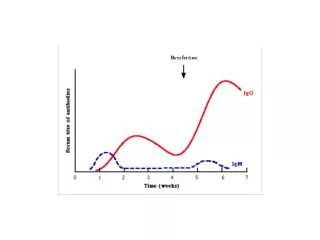
Sero Conversion • Seroconversion is the development of detectable specific antibodies to microorganisms in the blood serum as a result of infection or immunization.
Sero reversion • Seroreversion is the opposite of seroconversion. This is when the tests can no longer detect antibodies or antigens in a patient’s serum
Testing paired Samples • Testing for infectious diseases is performed on acute and convalescent specimens (about 2 weeks apart) Paired sample. • Must see 4-fold or 2-tube rise in titre to be clinically significant
Majority Diagnostic tests are Serological tests • There are several serology techniques that can be used depending on the antibodies being studied. These include: ELISA, agglutination, precipitation, complement-fixation, and fluorescent antibodies.
Antigen and Antibody reactions can be identified by different methods
Precipitation • Principle • Soluble antigen + antibody (in proper proportions) –> visible precipitate • Lattice formation (antigen binds with Fab sites of 2 antibodies) • Examples • Double diffusion (Ouchterlony) • Single diffusion (radial immunodiffusion) • Imunoelectrphoresis • Immunofixation
Agglutination • Principle • Particulate antigen + antibody –> clumping • Lattice formation (antigen binds with Fab sites of 2 antibodies forming bridges between antigens) • Examples • Direct agglutination (Blood Bank) • Passive Hemagglutination (treat RBC's with tannic acid to allow adsorption of protein antigens) • Passive latex agglutination (antigen attached to latex particle)
Neutralization reactions • Similar in principle and interpretation of results • Antibody-binding • Hemagglutination inhibition (serum antibody reacts with known nonparticulate antigen –> binding occurs) • Neutralization (antibody neutralizes toxin) • After binding, antibody is not available to react in indicator system • Results: • NO agglutination or NO hemolysis = positive reaction • Agglutination or hemolysis = negative reaction (antibody not bound in origin
Neutralization reactions • Generally, positive control samples used in inhibition or neutralization tests show no reaction and negative control samples show a reaction (opposite of results in direct agglutination testing) • Example of inhibition: Hemagglutination inhibition test for rubella • Example of neutralization: antistreptolysin O test (ASO)
Complement fixation (CF) • Antibody and antigen allowed to combine in presence of complement • If complement is fixed by specific antigen-antibody reaction, it will be unable to combine with indicator system • Precautions • Serum must be heat-activated • Stored serum becomes anti-complementary • Extensive QC/standardization required • Only use for IgM antibodies
A serum sample is electrophoresed through an agar medium. A trough is cut in the agar and filled with Ab. A precipitin arc is then formed. Because Ag diffuses radially and Ab from a trough diffuses, the reactants meet in optimal proportions for precipitation. Imunoelectrphoresis (IEP)Qualitative
Serology can be done on various speciemns • Some serological tests are not limited to blood serum, but can also be performed on other bodily fluids such as semen and saliva, which have (roughly) similar properties to serum. • Serological tests may also be used forensically, generally to link a perpetrator to a piece of evidence (e.g., linking a rapist to a semen sample).
Enzyme immunoassay (EIA/ELISA) • Sandwich technique” • Monoclonal or polyclonal antibody adsorbed on solid surface (bead or microtiter plate) • Add patient serum; if antigen is present in serum, it binds to antibody coated bead or plate • Add excess labelled antibody (antibody conjugate); forms antigen-antibody-labelled antibody “sandwich” (antibody in conjugate is directed against another epitope of antigen being tested) • Add substrate, incubate, and read absorbance • Washing required between each step • Absorbance is directly proportional to antigen concentration
ELISA methods takes over • Enzyme-linked immunosorbent assay, also called ELISA, enzyme immunoassay or EIA, is a biochemical technique used mainly in immunology to detect the presence of an antibody or an antigen in a sample. The ELISA has been used as a diagnostic tool in medicine • Because the ELISA can be performed to evaluate either the presence of antigen or the presence of antibody in a sample
ELISA Most popular technological advance in Laboratory Medicine • ELISA methods can detect any infectious disease provided if we have antibodies and antigen to any infection, enzyme or any substance
Serology applications in.. • HIV testing • Serum HCG (pregnancy) • Tests for hepatitis antigens and antibodies • Antibodies to bacteria • Hepatitis Serology
Nephelometry • Procedure • Serum substance reacts with specific antisera and forms insoluble complexes • Light is passed through suspension • Scattered (reflected) light is proportional to number of insoluble complexes; compare to standards • Examples • Complement component concentration • Antibody concentration (IgG, IgM, IgA, etc.) • Immunofluorescence
Immunofluorescence • Direct – add fluorescein-labeled antibody to patient tissue, wash, and examine under fluorescent microscope • Indirect – add patient serum to tissue containing known antigen, wash, add labeled antiglobulin, wash, and examine under fluorescent microscope • Examples • Testing for Antinuclear Antibodies (ANA) • Fluorescent Treponemal Antibody Test (FTA-Abs)
Fluorescence polarization immunoassay (FPIA) • Principle • Add reagent antibody and fluorescent-tagged antigen to patient serum • Positive test • Antigen present in patient serum binds to reagent leaving most tagged antigen unbound • Unbound labeled antigens rotate quickly reducing amount of polarized light produced • Negative test • If no antigen present in patient serum, tagged antigen binds to reagent antibody • Tagged antigen-antibody complexes rotate slowly giving off increased polarized light
Flow cytometry • Method of choice for T- and B-cell analysis (lymphocyte phenotyping) • Principle • Incubate specimen with 1 or 2 monoclonal antibodies tagged with fluorochrome • Single cells pass through incident light of instrument (laser) which excites fluorochrome and results in emitted light of different wavelength • Intensity of fluorescence measured to detect cells possessing surface markers for the specific monoclonal antibodies that were employed • Forward light scatter indicates cell size or volume • 90° side-scattered light indicates granula
Common uses Flow cytometry • DNA analysis • Reticulocyte counts • Leukaemia/lymphoma classification • CD 4 cell estimations in AIDS/HIV patients.
Other Applications of agglutination tests in Serology i. Determination of blood types or antibodies to blood group antigens. ii. To assess bacterial infections e.g. A rise in titer of an antibody to a particular bacterium indicates an infection with that bacterial type. N.B. a fourfold rise in titer is generally taken as a significant rise in antibody titer.
Georges-Fernand-Isidor Widal • Widal in 1896, and Widal & Sicard in 1896 described the Widal reaction, and this test has proved of value in cases where positive cultures have been unobtainable
Widal test a Popular test in diagnosis of Typhoid Fever • The Widal test is a presumptive serological test for Enteric fever or Undulant fever. In case of Salmonella infections, it is a demonstration of agglutinating antibodies against antigens O-somatic and H-flagellar in the blood.
Widal test is century old ,Is it loosing importance ? • In this reaction antibodies react with antigens on the surface of particulate objects and cause the objects to clump together, or agglutinate. These reactions were the earliest to be adapted to diagnostic laboratory. Widal test is used for diagnosis of typhoid fever. This test, developed by Georges Fernand I. Widal (French physician) in 1896, is now supplemented by more sophisticated procedures.
Widal test – A standard tube agglutination test • Test can be performed by the tube dilution technique which permits, the assay of antibody titre. In this, a constant amount of the antigen is added to a series of tubes containing serum dilutions. After mixing, the tubes are incubated at a particular temperature and the highest dilution of serum showing visible agglutination is determined.
Agglutination how it appear after reactivity • O agglutination is granular • H agglutination is loose and floccular
Principle of the Test • A classic example of the agglutination reaction is seen in the widal test for diagnosis of typhoid fever. In this test the antibody content of the patient's serum, is measured by adding a constant amount of antigen (Salmonella typhi) to the serially diluted serum.
Reading the Widal Test • Read the results by viewing the tubes under good light against the dark background with x2 magnifying lens • Do not shake tubes before reading the results • Read titers as greatest dilutions giving visible agglutinations. • Limiting agglutination is 1in 200 the titer is 200 not to be reported as 1/200.
Interpretation of Widal test • Test results need to be interpreted carefully in the light of past history of enteric fever, typhoid vaccination, general level of antibodies in the populations in endemic areas of the world.