Download
1 / 59
590 likes | 712 Views
An overview of nervous system development. How are all the different regions and cell types specified? How do they arise in the correct areas? How do all these regions/cell types get connected together?. Patterning, proliferation and neurogenesis. Specification of cellular identities.

E N D
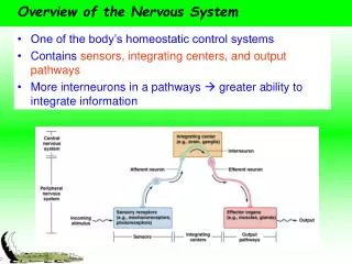
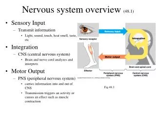
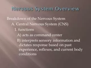
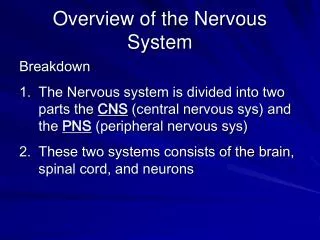
An overview of nervous system development How are all the different regions and cell types specified? How do they arise in the correct areas? How do all these regions/cell types get connected together?
Processes to consider: • Induction of the nervous system • Neurulation (formation of the neural tube) • Patterning of major axes • Proliferation • Establishment of cell fates • Cell migration • Axon guidance • Synaptogenesis • Cell death • Synaptic refinement • Myelination References: Jessell and Sanes (2000); Kandel, Jessell and Schwartz, Principles of Neuroscience
Clinical relevance • Birth defects • Psychiatric disorders • Regeneration • Stem cell therapeutics
Proliferation and Neurogenesis Amount of proliferation controlled by amount of asymmetric cell division When a progenitor cell divides does it make: - Two progenitors? - One progenitor and one neuron? - Two neurons?
Microcephaly • Small head size (small brain) • Moderate to severe mental retardation • Seizures (rare) • Genetically heterogeneous (six loci identified)
Chuas or “rat people” Many found at shrine to 17th century Sufi saint 1st cousin marriages - common in British Pakistani community too
Can the study of microcephaly tell us anything about control of proliferation and evolutionary expansion of the neocortex?
Principle of linkage analysis Recombination in meiosis: Variants near each other on the same chromosome (“linked”) tend to be inherited together. The co-inheritance of a neutral molecular marker with a disorder implies the mutant gene is near that marker.
MCPH5 mapped to ASPM gene Homologous to abnormal spindle (asp) gene in Drosophila Mutations lead to truncated protein Bond et al., (2002) Nature Genet. 32: 316
Expression of ASPM in developing mouse brain Ventricular zone
A number of other genes that cause Microcephaly • have also been identified: • MCPH1: Microcephalin - control of mitosis (Jackson et al., (2002) Am J Hum Genet. 71, 136-42) • MCPH3: CDK5RAP2 • MCPH6: CENPJ - both involved in chromosome segregation in mitosis (Bond et al., (2005) Nat Genet. 37, 353-5)
How do mutations in genes controlling mitosis lead to microcephaly? Aspm mRNA expressed at early stages: - Divisions are symmetric - Progenitor pool expanding Aspm mRNA downregluated at later stages: - Divisions are asymmetric - Neurons being generated
Symmetric divisions at early stages generate two neuroepithelial progenitors - expand pool of progenitors Asymmetric divisions at later stages generate one postmitotic neuron and one progenitor - as each progenitor can only generate a limited number of neurons this eventually depletes pool of progenitors and leads to fewer neurons
Asymmetric distribution of cytoplasmic factors coordinated with orientation of mitotic spindle
Knockdown of Aspm results in more asymmetric divisions at early stages Effect is more progeny adopt neuronal fate and fewer retain neuroepithelial progenitor fate
Mutation of Aspm (or other genes implicated in microcephaly) causes: Defect in alignment of mitotic spindle with axis of cell Increase in asymmetric division at early stages Failure to expand progenitor pool Premature generation of neurons Reduction in brain size
Conclusions: • Microcephaly caused by mutations in many genes • All involved in mitosis somehow • Defects in Aspm affect symmetric division • Progenitor pool fails to expand - depleted too early • Small brain results • ASPM, MCPH1, CDK5RAP2 all show evidence of positive selection in lineage leading to humans • Inference: Mutations in these genes that increased brain size may have been selected for in human lineage
Diversity of cell types and functions Red blood cells Hair cells in cochlea Cardiac muscle cells Nerve cells Skin cells
What makes cells different is they make different proteins Some proteins made only in specific cell types: e.g., hemoglobin, insulin
Each tissue/cell type has a different profile • - Express different genes related to their specific functions • (neurotransmitter receptors, ion channels, etc.) • Express specific code of transcription • factors that control expression of all the • other genes that make each cell unique • (i.e. that specify its “identity”) • How do they come to express that • spectrum of transcription factors?
Process of reiterative subdivision of embryo and progressive restriction of potential. - specification of intermediate fates of dividing cells en route to specification of final fates of postmitotic cells Occurs through series of cellular interactions beginning at the first cell division and continuing throughout development as morphogenetic movements shape embryo.
Patterning and establishment of cell fates 1. Gradients of diffusible molecules specify different fates at different concentrations 2. Interactions between neighbouring cells also influence cell fates
Different neuronal types generated from specific progenitor pools
Progenitor pools are specified by code of transcription factors (Briscoe et al., 2000)
Floor plate of spinal cord can induce ectopic motorneurons motoneurons Floor plate Floor plate grafted Wild-type situation Floor plate ablated (Embryological experiments in chick)
Sonic hedgehog is a secreted protein expressed in floor plate Shh conc.
How do you get such sharp borders? Cross-repression between transcription factors:
Cross-repression: • Nkx2.2 activates its own transcription and represses Pax6 • Pax6 activates its own transcription and represses Nkx2.2 • Both genes can’t be expressed in same cell - slight imbalance amplified - graded expression becomes sharp - individual cells specified as one fate or another
Combinatorial code of transcription factors • Control expression of other genes (i.e., turn on whole “profile” of gene expression for different subtypes of neurons) • These downstream “effector” genes control various aspects of cell fate: • Connectivity • Neurotransmitter expression • Expression of ion channels/receptors, etc.
Mutations in Shh lead to Holoprosencephaly (OMIM: 142945)
Specification of clinically important cell types Midbrain dopaminergic neurons degenerate in Parkinson’s disease
Parkinson’s disease Primary symptoms: • Tremor: an uncontrollable trembling or shaking • Rigidity: an abnormal stiffness of the muscles • Bradykinesia: an extreme slowness of movement and reflexes. Caused by progressive loss of midbrain dopaminergic neurons - can be familial (often early-onset) Current therapies (L-dopa) only moderately effective