Download
1 / 43
430 likes | 608 Views
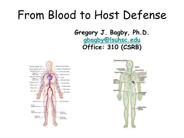
From Blood to Host Defense. Gregory J. Bagby, Ph.D. gbagby@lsuhsc.edu Office: 310 (CSRB ). From Blood to Host Defense. Blood Components and function Hemostasis and clotting The host defense system Innate immune system Pathogen recognition Inflammatory response

E N D
From Blood to Host Defense Gregory J. Bagby, Ph.D. gbagby@lsuhsc.edu Office: 310 (CSRB)
From Blood to Host Defense • Blood • Components and function • Hemostasis and clotting • The host defense system • Innate immune system • Pathogen recognition • Inflammatory response • Local to systemic responses and integration • Adaptive immune system • Humoral immune system and antibodies • Cell-mediated immune system
Liver Bone marrow Vascular system Lymphoid system The Relationship between Blood and Host Defense Lymph node
The Relationship between Blood and Host Defense • Cellular elements of blood and the immune system produced and/or originate in bone marrow via hematopoiesis • Red blood cells (erythrocytes) –important in O2 and CO2 transport. • White blood cells (leukocytes) – key roles in host defense. • Platelets – perform role in hemostasis and clotting • Plasma proteins – many produced in liver or by cells of the host defense system
From Blood to Host Defense Blood Components and Function Gregory J. Bagby, Ph.D. gbagby@lsuhsc.edu Office: 310 (CSRB)
Blood Components and Function • The components of blood • Assessment of Cell Numbers and Types • Regulation of hematopoiesis • Regulation of erythrocyte production
What is blood? • Blood is a fluid that normally circulates through the lumen of the cardiovascular system (heart, arteries, capillaries, and veins) • Two major components • Plasma – liquid (a complex solution) • Formed elements (cells and cell fragments)
What are the main functions of blood? • Transport water, ions, nutrients and waste products to and from tissues • Ions – sodium, chloride, calcium, bicarbonate, etc • Nutrients – glucose, amino acids, lipids, oxygen • Waste products – urea, lactic acid, carbon dioxide • Transport signaling molecules (hormones) from cells of origin to target cells • Defending or protecting the blood and extravascular compartments • Hemostasis and clotting • Role in host defense - means by which elements of the host defense system travel to the thymus, mucosal tissues, liver, lymphoid tissue, and sites of tissue injury or infection.
Regulation of Blood Cell Production The Components of Blood • Plasma – liquid component • Formed elements 1. Erythrocytes 2. Leukocytes 3. Platelets These components are maintained within a narrow range (concentration or counts/ml), but new replace old. Production and/or removal of each constituent if regulated to maintain homeostasis.
Hematocrit or PCV Plasma includes water, ions, proteins, nutrients, hormones, wastes, etc. Normal values Men = 45% Women = 42% Buffy coat of leukocytes separates RBC from plasma. The hematocrit is a rapid assessment of blood composition. It is the percent of the blood volume that is composed of RBCs (red blood cells).
How Much Blood Is in the Body? • Blood volume normally 8% of body weight • Blood volume = 5.6 L in 70 kg man • Erythrocyte Volume (45% hematocrit): 0.45 x 5.6 L = 2.5 L • Plasma Volume: 5.6 L – 2.5 L = 3.1 L
(Liver) (Immune) (Liver) Constituents of Plasma
Constituents of Plasma (continued) Bound to albumin or in lipoproteins
Serum and Plasma • Serum is plasma with fibrinogen and other proteins involved in clotting removed as a result of clotting. • Serum is often used for analysis instead of plasma • Need anticoagulant to obtain plasma • Fewer interfering substances in serum (less protein)
5,000,000/mm3 250,000/mm3 7,000/mm3 Suspended Formed Elements The major forms of “cells” in the blood. Among these, only the leukocytes are true cells with nuclei.
Normal Range of Blood Cell Numbers (counts/liter) Oxford Handbook of Clin Med and Anatomy & Physiology in Health and Illness (Ross and Wilson)
Carry O2 from the lungs / CO2 to the lungs Contain large amounts of hemoglobin (35% of mass) Men: 16 g / 100 ml Women: 14 g / 100 ml Biconcaved discs (high surface:volume ratio to maximize diffusion capacity. Aids in flow through small vessels.) Red Blood Cells: Erythrocytes
Polymorphonuclear (PMN) leukocytes (granulocytes) Leukocytes and Platelets • Cells of the Immune System • Small percentage of total blood cells • Types: • Neutrophils (50-70%) • Eosinophils (1-4%) • Basophils (0.1%) • Monocytes (2-8%) • Lymphocytes (20-40%) • Platelets (250,000 per mm3 of blood)
Blood Components and Function • The components of blood • Assessment of Cell Numbers and Types • Regulation of hematopoiesis • Regulation of erythrocyte production
Assessment of Cell Numbers and Types • Manual counts using a microscope • Morphology/staining • Auto analyzers such as a Coulter Counter • Morphology • Flow cytometry • Morphology/Specific Antibody binding to antigens on/in cells (proteins)
Manual Blood Cell Count Determination Using a Light Microscope, Hemacytometer and Blood Smear
RBC red blood cell (count 10^6 cell/microliter) Hgbhemaglobin (gm/dl) Hct hematocrit (%) Rtcreticulocytes Mcv mean corpuscular volume (fl) (femtoliters) -- normal R.m indoor adult males 72-76 --- normal adult humans 86-98 Mch mean cell hemaglobin (pg) - normal R.m. indoor adult males 21.8-24.6 --- normal adult humans 27-32 Mchc mean cell hemaglobin concentration(%) - normal R.m. indoor adult males 29.6-31.2 --- normal adult humans 32-36 Rdw red cell distibution width --- normal adult males 11-15 WBC white blood cell (count 10^3/microcliiter) Sgssegmented neutrophils(%) Bnd banded neutrophils (%) Eos eosenophils (%) Bsobasophils (%) Mnomoncytes (%) Lym lymphocytes (%) Plt platelets (count/microliter) Auto Analyzyer – Coulter Counter
Flow Cytometry of Fluorescence Activated Cell Sorting (FACS) Detection Laser FALS Sensor Fluorescence detectorScattered light detector - + Fluorescence labeled antibodies against specific proteins Charged Plates Single cells sorted into test tubes Modified from Purdue University Cytometry Laboratories
Laser FALS Sensor Forward Angle Light Scatter (Forward Scatter) When a cell intercepts the laser beam, the light scattered in the forward direction (along the same axis that the laser light is traveling) is detected in the forward scatter channel. Forward scatter Size and shape of cell – bigger the shadow the bigger the cell
Laser FALS Sensor 90LS Sensor 90 Degree Light Scatter (Side Scatter) The amount of light scattered to the side (perpendicular to the axis that the laser light is traveling) is detected in the side or 90o scatter channel. Forward scatter Reflected light Shape (irregular) and optical heterogeneity of cells – granulation (# of organelles increases side scatter) Side scatter
Scale associated with # of events or cells 1000 200 100 50 40 30 20 15 8 Light Scatter Gating Neutrophils Forward scatter Monocytes Lymphocytes 200 400 600 800 1000 0 Side Scatter Modified from Purdue University Cytometry Laboratories
Detection of Fluorescence The amount of fluorescence is detected in the side or 90o scatter channel. Laser Excitation FALS Sensor Forward scatter Emissionintensity Fluorescence detector Detection of protein on surface or inside cell by binding of fluorochrome-conjugated antibody (Phenotype or function).
Scale associated with # of events or cells 1000 200 100 50 40 30 20 15 8 Light Scatter Gating Neutrophils Forward scatter Monocytes Lymphocytes 200 400 600 800 1000 0 Side Scatter Modified from Purdue University Cytometry Laboratories
Gating on Lymphocytes and Detecting CD3+ Cells that are either CD4+ or CD8+ CD3+ (T lymphocytes) 1 2 45% 2% Log PE Fluorescence (CD4) 3 4 27% 26% .1 1 10 100 1000 Log FITC Fluorescence (CD8)
CD4 CD8 CD3 CD3 CD # = cluster designation number CD20 CD3+CD4+ CD3+CD8+ (Th1 cell) (Antigen specific CTL) T helper cell Cytotoxic T cell B cell CD3-CD20+ Immunophenotyping of Lymphocytes IFNgamma IFNgamma
Stem cells in the bone marrow constitute an important precursor of many of the formed components in the blood.
Hematopoietic Growth Factors HGF:receptor binding activates intracellular signaling cascades
Erythropoietin Colony-Stimulating Factors Interleukins Thrombopoietin Stem Cell Factor Others (TNF, Interferons) Erythrocytes Granulocytes and monocytes Various Leukocytes Platelets Many Blood types Name: Product: Major Hematopoietic Growth Factors: (Derived from Table 14-4)
Erythrocytes • Produced in the bone marrow • Lose nuclei and organelles • Life-span = 120 days • 250 billion cells made per day • Destroyed in liver and spleen • Bilirubin is the breakdown product (gives plasma its color) • Erythrocyte production tightly regulated by hormones.
Erythropoiesis is hormonally regulated: decreased oxygen delivery to the kidney causes the secretion of erythropoietin, which activates receptors in bone marrow, leading to an increase in the rate of erythropoiesis. • Erythropoietin Used Clinically: • Blood loss • Renal failure • In conjunction with chemotherapy
Erythrocytes • Produced in the bone marrow • Lose nuclei and organelles • Life-span = 120 days • 250 billion cells made per day • Destroyed in liver and spleen • Bilirubin is the breakdown product (gives plasma its color) • Erythrocyte production tightly regulated by hormones. • Erythrocyte production dependent on folic acid, vitamin B12 and iron
Folic Acid and Vitamin B12 Folic acid • A vitamin found in leafy plants, yeast, and liver • Is required for synthesis of the nucleotide base thymine • Essential for the formation of DNA and normal cell division Vitamin B12 • Is found only in animal products- Strict vegetarian diets are often deficient in B12 • Absorption in GI tract requires “intrinsic factor” • Is required for the action of folic acid (DNA replication)
Iron Metabolism 50% The availability of dietary iron can be a limiting factor in rbc production, so storage and recycling mechanisms are highly developed in humans as a protection from anemia. 25% Transferrin Balance input vs output (95% recycled). Maintaining iron balance important for adequate hemoglobin/rbc production. Iron deficiency – anemia Hemochromatosis –iron toxicity 25% Ferritin
Anemia and Polycythemia • Anemia: Decreased ability of blood to carry oxygen - Decreased # erythrocytes - Decreased concentration or performance of hemoglobin within erythrocytes - Combination of both • Polycythemia: More erythrocytes than normal (opposite of anemia) - Increased viscosity of blood - altitude
Sickle-cell anemia • Genetic mutation alters one amino acid in hemoglobin • Fiber-like structures form during low [O2], distorting erythrocyte into sickle shape • Capillary blockage • Tissue damage • Destruction of deformed erythrocyte • Anemia
Regulation of Total Blood Cell production • All blood cells are derived from multipotent/pluripotenthematopoietic stem cells • Differentiation and proliferation of stem cells (the path taken) is stimulated by hematopoietic growth factors(HGF’s).