Download
1 / 26
260 likes | 358 Views
200 µm. LE 12-2b. Growth and development. 20 µm. LE 12-2c. Tissue renewal. Types of Cells. Proliferation Differentiation Highly differentiated cells: Quiescent cells: Highly mitotic cells:. How to determine the cycle?. Experiment 1. S. G 1. LE 12-13a. S. S.

E N D
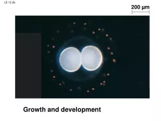
200 µm LE 12-2b Growth and development
20 µm LE 12-2c Tissue renewal
Types of Cells • Proliferation Differentiation • Highly differentiated cells: • Quiescent cells: • Highly mitotic cells:
Experiment 1 S G1 LE 12-13a S S When a cell in the S phase was fused with a cell in G1, the G1 cell immediately entered the S phase—DNA was synthesized.
Experiment 2 M G1 LE 12-13b M M When a cell in the M phase was fused with a cell in G1, the G1 cell immediately began mitosis—a spindle formed and chromatin condensed, even though the chromosome had not been duplicated.
Mitosis and Cancer • Common and severe • Some form strikes 1/3; 20% death; 20% health care costs • Not a single disease • Early detection and treatment is vital invade neighboring tissue neoplasm tumor cell division metastasis
Cells anchor to dish surface and divide (anchorage dependence). When cells have formed a complete single layer, they stop dividing (density-dependent inhibition). LE 12-18a If some cells are scraped away, the remaining cells divide to fill the gap and then stop (density-dependent inhibition). 25 µm Normal mammalian cells
Cancer cells do not exhibit anchorage dependence or density-dependent inhibition. LE 12-18b 25 µm Cancer cells
The genetic nature of cancer • The clonal nature of cancer • Analyzing the cancer and normal cells • Most happen later in life • Proto-oncogenes and oncogenes • Recessive oncogenes and tumor suppressor genes
Tumor LE 12-19a Glandular tissue A tumor grows from a single cancer cell. Cancer cells invade neighboring tissue.
Lymph vessel LE 12-19b Blood vessel Metastatic tumor Cancer cell A small percentage of cancer cells may survive and establish a new tumor in another part of the body. Cancer cells spread through lymph and blood vessels to other parts of the body.
Searching for Oncogenes • Cell cycle regulatory genes • A lot of cancer are associated with DNA virus • Some cancers are familial • In vitro mutagenesis on cultured cells • Loss of heterozygocity
Some Oncogenes • Both viral AND cellular • src (Rous sarcoma v.): Prototype of a family of membrane associated tyrosine kinases • over expressed or activated in cancer Kinases: abl (abelson murine leukemia v.) trk (human colon carcinoma v.) GTPase: ras (rat sarcoma v.) Transcription factors: fos, myc (chicken sarcoma v.) EGF: erb A (avian erythroblastosis v.) EGF: epithelial PDGF: platelet - derived CSF: colony stimulating FGF: basic fibroblast
Some Familial Cancers • Rb • familial polyposis coli • Wilms Tumor • familial breast cancer • Li- Fraumeni familial cancer
EGF EGFR-P Quiescent Cell Division P53 p53-p P21 CDK2-p21 Cyclin E CyclinA CDK2 ras fos Jun D-cyclin, CDK 2/4/6 Cyclin E Rb-p Rb Enzymes for DNA synthesis Rb E2F E2F G1 S
Figure 9.7 • Multiple nuclei • Lost adhesion • Irregular cell shape • Vessel invation • Therapy: • Vessel growth inhibitor • Nucleotide analog
Protection against cancer: • Apoptosis • Immunity • Telomeres
Cancer Protection • Immunity • Apoptosis ras mutation 5q gene loss ch 18 loss increased growth adenoma III adenoma II adenoma I normal other ch ch 17 carcinoma metastasis p53


![[12-2B] Box & Whisker Plots](https://cdn3.slideserve.com/6306071/slide1-dt.jpg)