Download
1 / 56
780 likes | 1.49k Views
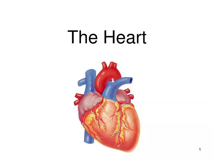
The Heart. Functions of the Heart. Generating blood pressure Routing blood separates pulmonary and systemic circulations Ensuring one-way blood flow: valves Regulating blood supply Changes in contraction rate and force match blood delivery to changing metabolic needs.

E N D
Functions of the Heart • Generating blood pressure • Routing blood • separates pulmonary and systemic circulations • Ensuring one-way blood flow: valves • Regulating blood supply • Changes in contraction rate and force match blood delivery to changing metabolic needs
Heart’s position in thorax • In mediastinum – behind sternum and pointing left, lying on the diaphragm • It weighs 250-350 gm (about 1 pound) Feel your heart beat at apex (this is of a person lying down)
Chest x rays Lateral (male) Normal female
Layers of the heart wall • Muscle of the heart has an inner and an outer membrane covering • The layers from out to in: • Epicardium = epithelial tissue/visceral layer of serous pericardium • Myocardium = the muscle • Endocardium= epithelial tissue lining the chambers
Pericardium(see next slide) Starting from the outside… Without most of pericardial layers
Coverings of the heart: pericardium Three layered: • Fibrous pericardium (anchors the heart in place) • Serous pericardium of layers: two parts • Parietal layer- Additional insulation • Visceral layer (aka epicardium)- On the heart/part of its wall; made of muscle (Between the layers is pericardial cavity w/ pericardial fluid)
(Visceral Pericardium) Epicardium Pericardium Myocardium
Two circuits • Pulmonary circuit • blood to and from the lungs • Systemic circuit • blood to and from the rest of the body *Vessels carry the blood through the circuits • Arteries carry blood away from the heart • Veins carry blood to the heart • Capillaries permit exchange
The heart=a muscular double pump with 2 functions Overview • Right Side: • Receives oxygen-poor blood from the body • Pumps it to the lungs to pick up O2and dispel CO2 • Left Side: • Receives oxygenated blood returning from the lungs • Pumps oxygen rich blood to the body to supply oxygen and nutrients to the body tissues
Heart Chambers vs. Valves • Structural Differences in heart chambers • Chambers= atria and ventricles • The left side = more muscular than the right side • Functions of valves • Atrioventricular (AV) valves prevent backflow of blood from the ventricles to the atria • Semilunar valves prevent backflow into the ventricles from the pulmonary trunk and aorta
Chambers of the heartsides are labeled in reference to the patient facing you • Two atria • Right atrium • Left atrium • Two ventricles • Right ventricle • Left ventricle --------------------------------------------------------------------------------
Chambers of the heartdivided by septae: • Two atria-divided by interatrial septum • Right atrium • Left atrium • Two ventricles-divided by interventricular septum • Right ventricle • Left ventricle
Right Side • Superior Vena Cava- oxygen poor blood from the upper body returns to the heart • Inferior Vena Cava- oxygen poor blood from the upper body returns to the heart • Right Atrium- collects oxygen poor blood from the body; forces it through the tricuspid valve (AV) into the right ventricle • Right Ventricle- collects oxygen poor blood from the right atrium; forces it through the pulmonary valve (semilunar) into the lungs * Pulmonary arteries (2) carry blood to the lungs
W2L: Thickness of muscular walls The left ventricle is thicker than the right ventricle. Use what you already know about the heart to explain why there is such a difference.
W2L : ANSWER LV thicker than RV because it forces blood out against more resistance; the systemic circulation is much longer than the pulmonary circulation. Atria are thin because ventricular filling is done by gravity, requiring little atrial effort
Left Side * Pulmonary veins (4) carry oxygen rich blood from the lungs to the heart • Left Atrium- collects oxygen rich blood from the lungs; forces it through the mitral valve (AV)into the left ventricle • Left Ventricle- collects oxygen rich blood from the left atrium; forces it through the aortic valve (Semilunar) into the body • Is the largest/strongest chamber in the heart * Aorta (3) carry blood to the body
Importance of Valves • Valves open and close in response to pressure differences • Note papillary muscles, trabeculaecarnae & chordaetendinae (heart strings): keep valves from prolapsing (purpose of valve = 1 way flow)
Two for Two! Heart Tissue Model! Heart Flow! Create a flow chart of blood flow through the heart. Color code it! • Use the materials provided to represent the layers of the heart. • Be creative!!!!
To Do: • Go over heart dissection • Present Tissue Layers of the Heart • Graphic Organizer on Blood Flow • Cardiac Muscle
Bell Work • What does deoxygenated blood travel through when it leaves the heart to the lungs? • What does blood travel through when it returns oxygenated blood back to the heart from the lungs?
Bell Work Describe some characteristics of cardiac muscle (2-3).
Cardiac Muscle • Cell Characteristics: • Elongated and branching • Contains 1-2 centrally located nuclei • Contains actin and myosin myofilaments(striated look due to sarcomeres) • Vol.= 30% mitochondria (vs. 2% in skeletal muscle) • Intercalated disks: • Cell membranes interlock held together by desmosomes • Gap junctions allow action potentials to move from one cell to the next.
Heart Rhythm • Cardiac muscle cells can contract spontaneously and independently • Regulation of heart activity: • Autonomic nervous system • Epinephrine, thyroxine: heart rate • Low Ca2+ levels: heart rate • Intrinsic conduction system • Built into heart tissue & sets basic rhythm • Pacemaker = Sinoatrial (SA) Node
The Heartbeat • The Conducting System • Initiates and spreads electrical impulses in the heart • Two types of cells • Nodal cells • Pacemaker cells • Reach threshold first (AP) • Set heart rate • Conducting cells • Distributes stimuli to myocardium
The Heartbeat • The Conducting System (cont’d) • Heart is self-exciting • Pacemaker cells establish heart rate • Normal pacemaker is sinoatrial (SA) node • Impulse spreads from SA node: • Across atria • To atrioventricular (AV) node- causes contraction of atria • To AV bundle and bundle branches • Via Purkinje fibers to ventricles- causes contraction of the ventricles
SA node activity and atrial activation begin. SA node Time = 0 Stimulus spreads across the atrial surfaces and reaches the AV node. AV node Elapsed time = 50 msec There is a 100-msec delay at the AV node. Atrial contraction begins. AV bundle Bundle branches Elapsed time = 150 msec The impulse travels along the interventricular septum within the AV bundle and the bundle branches to the Purkinje fibers. Elapsed time = 175 msec The impulse is distributed by Purkinje fibers and relayed throughout the ventricular myocardium. Atrial contraction is completed, and ventricular contraction begins. Purkinje fibers Elapsed time = 225 msec
Cardiac Muscle Contraction • Heart muscle: • Is stimulated by nerves and is self-excitable (automaticity) • Contracts as a unit; no motor units • Cardiac muscle contraction is similar to skeletal muscle contraction, i.e., sliding-filaments
Differences Between Skeletal and Cardiac Muscle Physiology • Action Potential • Cardiac: Action potentials conducted from cell to cell. • Skeletal, action potential conducted along length of single fiber • Rate of Action Potential Propagation • Slow in cardiac muscle b/c of gap junctions and small diameter of fibers. • Faster in skeletal muscle due to larger diameter fibers.
Heartbeat • Def.- a two-part pumping action that takes about a second • Diastole- filling of the ventricles • Blood collects in the in the upper chambers (the right and left atria) • Atria contract forcing blood through the tricuspid &mitral valves into the resting lower chambers (the right and left ventricles) • Systole- contraction of the ventricles • Begins when the ventricles fill with blood • Ventricles contract forcing pulmonary and aortic valves open • Right ventricle to the lungs & left ventricle to the heart
Heartbeat • Normal rate: 60-100 beats • Slow: bradycardia • Fast: tachycardia
Heart sounds • Called S1 and S2 • S1 is the closing of AV (Mitral and Tricuspid) valves • S2 is the closing of the semilunar (Aortic and Pulmonary) valves • Murmurs: the sound of flow • Can be normal • Can be abnormal
Places to auscultate: Routine places are at right and left sternal border and at apex
Homework • Research three types of heart disease and include the following: • Definition • Causes • Symptoms • Prevention/treatment
Autonomic innervation • Sympathetic • Increases rate and force of contractions • Parasympathetic (branches of Vagus n.) • Slows the heart rate For a show on depolarization: http://education.med.nyu.edu/courses/old/physiology/courseware/ekg_pt1/EKGseq.html
Note that the usual name for “anterior interventricular artery” is the LAD (left anterior descending)
ANTERIOR VIEW Each atrium has an “auricle,” an ear-like flap