Download
1 / 24

E N D
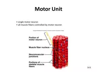
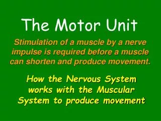
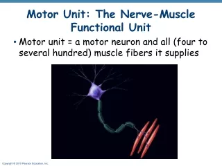
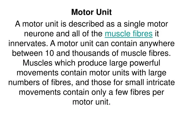
Motor Unit A motor unit is described as a single motor neurone and all of the muscle fibres it innervates. A motor unit can contain anywhere between 10 and thousands of muscle fibres. Muscles which produce large powerful movements contain motor units with large numbers of fibres, and those for small intricate movements contain only a few fibres per motor unit.
Where the synaptic knobs of the neurone meet the muscle fibres is known as the neuromuscular junction. When an impulse reaches the neuromuscular junction, a neurotransmitter called Acetylcholine is released which filters across the synaptic cleft (microscopic space between the synaptic knob and motor end plate). This causes depolarisation of the motor end plate and puts the sliding filament theory of muscular contraction into practice.
The 'all or none' law as mentioned above also applies to the contraction of fibres within a motor unit. When a motor unit activates, all of the fibres within the unit contract and at full force, there is no strong or weak contraction. The strength of the resultant whole muscular contraction depends upon the number of motor units recruited
Another way of increasing the stength of a muscle contraction is by decreasing the time between impulses so that the muscle fibres do not have time to relax, resulting in a continuous wave of contractions known as wave summation. To produce a strong contraction all motor units in the muscle are recruited, but only for a short time. In order to increase the length of a contraction a kind of rotation system is implemented whereby some units contract while others rest and continuously alternate. This is known as spatial summation or tetanus
SPATIAL SUMMATION WAVE SUMMATION
Skeletal Muscle StructureAlthough skeletal muscles come in different shapes and sizes the main structure of a skeletal muscle remains the same. If you were to take one whole muscle and cut through it, you would find the muscle is covered in a layer of connective tissue known as the Epimysium. The Epimysium protects the muscle from friction against other muscles and bones. A large strong muscle, such as thoses forming your Quadriceps would have a large number of fibres within each bundle. A smaller muscle used for precision movement, such as those in the hand would contain far fewer fibres per Fasciculi.
Looking at each muscle fibre in detail, you can see they too are covered in a fibrous connective tissue, known as Endomysium which insulates each muscle fibre. Muscle fibres can range from 10 to 80 micrometers in diameter and may be up to 35cm long. Beneath the Endomysium and surrounding the muscle fibre is the Sarcolemma which is the fibres cell membrane and beneath this is the Sarcoplasm, which is the cells cytoplasm, a gelatinous fluid which fills most cells. This contains Glycogen and Fats for energy and also Mitochondria which are the cells powerhouses, inside which the cells energy is produced
Each muscle fibre itself contains cylindrical organelles known as Myofibrils. Each muscle fibre contains hundreds to thousands of Myofibrils. These are bundles of Actin and Myosin proteins which run the length of the muscle fibre and are important in muscle contraction. • Surrounding the Myofibril there is a network of tubules and channels called the Sarcoplasmic Reticulum in which Calcium is stored which is important in muscle contraction. Transverse tubules pass inwards from the Sacrolemma throughout the Myofibril, through which nerve impulses travel. • Each Myofibril can then be broken down into functional repeating segments called Sarcomeres.
SARCOMERE Sliding filament model of muscle contraction The sarcomeres are what give skeletal and cardiac muscles their striated appearance. • A sarcomere is defined as the segment between two neighbouring Z-lines (or Z-discs, or Z bodies). In electron micrographs of cross striated muscle the Z-line (from the German "Zwischenscheibe", the band in between the I bands) appears as a series of dark lines. • Surrounding the Z-line is the region of the I-band (for isotropic). • Following the I-band is the A-band (for anisotropic). Named for their properties under a polarizing microscope. • Within the A-band is a paler region called the H-band (from the German "Heller", bright). Named for their properties under a polarization microscope. • Finally, inside the H-zone is a thin M-line (from the German "Mittel", middle of the sarcomere). what give skeletal and cardiac muscles their striated appearance.
SARCOMERE - basic repeat unit of striated muscle, delimited by Z-linesI band - "paler zone" around Z-line (Isotropic - passes light in all directions)A band - "dark region" in center of sarcomere (Anisotropic - in different directions)M line - mid point of the sarcomereH zone - "paler zone" in the center of sarcomere around M line
some definitions related to muscle contraction... Summation - a 2nd contraction before 1st subsides fig* (cause is time differential of nerve/muscle) Tetany - sustained contractions (requires energy - ATP) Fatigue - under repeat stimulation, contractions get feebler, lactate accumulates, pH changes lead to stoppage of contractions Shivers - involuntary-summed muscle contractions which release waste heat, that warms body
Muscle Cell Proteins [4 types involved in contraction cycle] 1. THICK FILAMENT (A band)myosin* - 6 polypeptides twisted to form 2 helical fibers with globular ends, which have ATPase activity & an affinity to bind to actin myosins are... Molecular Motors&kinesin animations2. THIN FILAMENT (I band)G-actin* - globular protein which polymerizes into polymeric fiber... each globular actin unit contains a myosin binding site 3. Tropomyosin* - fiber-like protein which wraps helically around thin filament 4. Troponin - globular protein complex which binds Ca+2 & initiates contraction cycle is complex of 3 proteins, TroponinsC, I, & T, which bind Ca; Troponin C (18 kD) binds Ca reversibly.TnC binds TnI (23 kD) & TnT (37 kD), which change conformation in response to TC binding Ca.
Muscles can not push, they may only only CONTRACT (shorter via a pull) A muscle contraction is called a muscle TWITCH