Download
1 / 40
410 likes | 1.09k Views
OAP FLASH PAR DYSFONCTION DIASTOLIQUE. Mattéo Miquet DES Anesthésie-réanimation Desc Réanimation médicale Grenoble Mai 2006. Dysfonction diastolique ventriculaire. Définition : altération du remplissage ventriculaire avec une fraction d’éjection (FE) normale (>50%).

E N D
OAP FLASH PAR DYSFONCTION DIASTOLIQUE Mattéo Miquet DES Anesthésie-réanimation Desc Réanimation médicale Grenoble Mai 2006
Dysfonction diastolique ventriculaire Définition : altération du remplissage ventriculaire avec une fraction d’éjection (FE) normale (>50%) Fréquence : 30 à 50 % des IC sont diastoliques pures = IC à fonction systolique conservée (patient en OAP avec FE normaux)
Physiologie cardiaque : diastole ventriculaire 4 phases : - relaxation isovolumétrique - remplissage rapide - remplissage lent - systole auriculaire
Comment expliquer l ’insuffisance cardiaque avec une fraction d ’éjection normale L ’IC est liée au bas débit cardiaque La FE ne prédit pas du débit cardiaque FE = Volume TéléSystolique (VST)/Volume TéléDiastolique (VTD)
Comment expliquer l ’insuffisance cardiaque avec une fraction d ’éjection normale Normal ICS ICD VTDV 120 250 85 VTSV 50 200 35 FE % 60 20 60 VSV 70 50 50 DC, L/min 4.2 3.0 3.0 DC, normal % 0 - 30 - 30
Comment expliquer l’insuffisance cardiaque avec une fraction d ’éjection normale En l ’absence de la mesure du volume télédiastolique ventriculaire gauche, la FEVG ne peut pas prédire le débit cardiaque
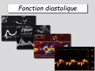
Diagnostic de la dysfonction ventriculaire diastolique Ne peut être porté par la seule clinique, l ’ECG ou la radiographie
Diagnostic de la dysfonction diastolique = examens complémentaires
OAP flash : circonstances 2 cadres : - un événement aigü : ischémie myocardique, HTA - évolution du processus chronique qui accompagne l’IC
Évolution du processus chronique qui accompagne l’IC activation du SRAA rétention hydrique
OAP : traitement • Ralentir la FC (éviter la tachycardie) • Préserver la systole auriculaire • Diurétique : attention à l’hypotension • Restriction sodée • Dérivés nitrés à action prolongée • IEC, ARAII • Traiter une poussée hypertensive • Traiter une ischémie myocardique
Conclusion Savoir diagnostiquer cette pathologie Nécessité d’une échographie cardiaque Pas de place pour la dobutamine
Etiologies de l ’insuffisance diastolique -cardiopathies ischémiques et hypertrophiques -causes extramyocardiques : péricardite, IVD, valvulopathies -vieillissement
Diastolic Heart Failure (EF>50%) Systolic Heart Failure (EF<50%) Symptoms Dyspnea on exertion 85 96 Paroxysmal nocturnal dyspnea 55 50 Orthopnea 60 73 Physical examination Jugular venous distension 35 46 Rales 72 70 Displaced apical impulse 50 60 S3 45 65 S4 45 66 Hepatomegaly 15 16 Edema 30 40 Chest radiograph Cardiomegaly 90 96 Pulmonary venous hypertensio 75 80perte Scan tableau 3 cahier anesthésio
Table 2. LVEF: Meaningless in Terms of Cardiac Output Without the Coexisting LVEDV* Variables Normal SHF DHF-1 DHF-2 SDf LVEDV, mL 120 250 100 85 200 LVESV, mL 50 200 50 35 130 LVSV, mL 70 50 50 50 70 HR, beats/min 60 60 60 60 60 LVEF, % 60 20 50 60 35 CO, L/min 4.2 3.0 3.0 3.0 4.2 CO, from normal, % 0 - 30 - 30 - 30 0
Table 1. Principles of Management of Diastolic Heart Failure* Goal Therapy Reduction of congestion Salt restriction Less than 2 g daily Diuretics Thiazides and loop diuretics ACEIs Enalapril Lisinopril ARBs Candesartan Losartan Maintenance of rate control ß-blockers Atenolol, metoprolol Calcium channel blockers Diltiazem, verapamil Conversion of atrial fibrillation Atrioventricular pacing Optimal management of hypertension Antihypertensive agents ß-blockers Calcium channel blockers Diuretics ACEIs ARBs Spironolactone Prevention and treatment of myocardial ischemia ß-blockers Atenolol, metoprolol Calcium channel blockers Diltiazem, verapamil Nitrates Isosorbide dinitrate Isosorbide mononitrate Revascularization Percutaneous transluminal coronary angioplasty, coronary artery bypass surgery