Download
1 / 43
430 likes | 469 Views
Explore the fascinating world of genetics, from the study of genes and chromosomes to the replication and expression of genetic information. Learn about DNA replication, transcription, and more in this comprehensive guide.

E N D
Terminology • Genetics: the study of what genes are, how they carry information, how information is expressed, and how genes are replicated • Gene: a segment of DNA that encodes a functional product, usually a protein • Chromosome: structure containing DNA that physically carries hereditary information; the chromosomes contain the genes • Genome: all the genetic information in a cell
Terminology • Genomics: the molecular study of genomes • Genotype: the genes of an organism • Phenotype: expression of the genes
Figure 8.1a A prokaryotic chromosome. Chromosome Insert Fig 8.1a Disrupted E. coli cell
Figure 8.2 The Flow of Genetic Information. Parent cell DNA replication expression recombination Insert Fig 8.2 Genetic information can be transferred between cells of the same generation. Genetic information is used within a cell to produce the proteins needed for the cell to function. Genetic information can be transferred between generations of cells. New combinations of genes Transcription Translation Daughter cells Cell metabolizes and grows Recombinant cell
DNA • Polymer of nucleotides: adenine, thymine, cytosine, and guanine • Double helix associated with proteins • “Backbone” is deoxyribose-phosphate • Strands are held together by hydrogen bonds between AT and CG • Strands are antiparallel
Figure 8.3b DNA replication. 5′ end 3′ end Insert Fig 8.3b 3′ end 5′ end The two strands of DNA are antiparallel. The sugar-phosphate backbone of one strand is upside down relative to the backbone of the other strand. Turn the book upside down to demonstrate this.
Figure 8.3a DNA replication. 5′ end 3′ end Parental strand Parental strand 1 1 The double helix of the parental DNA separates as weak hydrogen bonds between the nucleotides on opposite strands break in response to the action of replication enzymes. 1 Replication fork Insert Fig 8.3a 2 2 Hydrogen bonds form between new complementary nucleotides and each strand of the parental template to form new base pairs. 2 3 Enzymes catalyze the formation of sugar-phosphate bonds between sequential nucleotides on each resulting daughter strand. 3 Daughter strand 3′end Parental strand 5′ end Parental strand Daughter strand forming The replication fork
DNA Synthesis • DNA is copied by DNA polymerase • In the 5' 3' direction • Initiated by an RNA primer • Leading strand is synthesized continuously • Lagging strand is synthesized discontinuously • Okazaki fragments • RNA primers are removed and Okazaki fragments joined by a DNA polymerase and DNA ligase
Table 8.1 Important Enzymes in DNA Replication, Expression, and Repair Insert Table 8.1
Figure 8.5 A summary of events at the DNA replication fork. REPLICATION The leading strand is synthesized continuously by DNA polymerase. Proteins stabilize the unwound parental DNA. 3' DNA polymerase 5' Enzymes unwind the parental double helix. 1 Replication fork Insert Fig 8.5 RNA primer Primase 5' DNA polymerase 3' DNA ligase Okazaki fragment Parental strand 3' DNA polymerase 5' The lagging strand is synthesized discontinuously. Primase, an RNA polymerase, synthesizes a short RNA primer, which is then extended by DNA polymerase. DNA ligase joins the discontinuous fragments of the lagging strand. DNA polymerase digests RNA primer and replaces it with DNA.
Figure 8.6 Replication of bacterial DNA. Replication forks REPLICATION An E. coli chromosome in the process of replicating 1 Origin of replication Parental strand Daughter strand Replication fork Replication fork Termination of replication Bidirectional replication of a circular bacterial DNA molecule
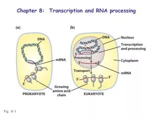
Transcription • DNA is transcribed to make RNA (mRNA, tRNA, and rRNA) • Transcription begins when RNA polymerase binds to the promoter sequence • Transcription proceeds in the 5' 3' direction • Transcription stops when it reaches theterminator sequence
Figure 8.7 The process of transcription. RNA polymerase DNA TRANSCRIPTION DNA mRNA RNA polymerase bound to DNA 1 Protein Promoter (gene begins) Template strand of DNA RNA polymerase RNA polymerase binds to the promoter, and DNA unwinds at the beginning of a gene. 1 Insert Fig 8.7 RNA is synthesized by complementary base pairing of free nucleotides with the nucleotide bases on the template strand of DNA. 2 RNA RNA nucleotides The site of synthesis moves along DNA; DNA that has been transcribed rewinds. 3 RNA synthesis Transcription reaches the terminator. 4 Terminator (gene ends) RNA and RNA polymerase are released, and the DNA helix re-forms. 5
Figure 8.11 RNA processing in eukaryotic cells. Intron Exon Intron Exon Exon RNA polymerase DNA Met Met In the nucleus, a gene composed of exons and introns is transcribed to RNA by RNA polymerase. DNA 1 Met RNA transcript Processing involves snRNPs in the nucleus to remove the intron-derived RNA and splice together the exon- derived RNA into mRNA. 2 1 Insert Fig 8.11 mRNA Met After further modification, the mature mRNA travels to the cytoplasm, where it directs protein synthesis. 3 Met Nucleus Cytoplasm
Translation • mRNA is translated in codons (three nucleotides) • Translation of mRNA begins at the start codon: AUG • Translation ends at nonsense codons: UAA, UAG, UGA
The Genetic Code • 64 sense codons on mRNA encode the 20 amino acids • The genetic code is degenerate • tRNA carries the complementary anticodon
Figure 8.8 The genetic code. Insert Fig 8.8
Figure 8.9.1-2 The process of translation. RNA polymerase TRANSLATION Ribosome Met DNA Leu DNA Met P Site mRNA Ribosomal subunit tRNA Protein Anticodon 1 Ribosomal subunit mRNA Start codon Second codon mRNA Components needed to begin translation come together. On the assembled ribosome, a tRNA carrying the first amino acid is paired with the start codon on the mRNA. The place where this first tRNA sits is called the P site. A tRNA carrying the second amino acid approaches. 1 2 Insert Fig 8.9 (1) and (2)
Figure 8.9.3-4 The process of translation. Peptide bond forms Met Met Phe DNA Leu Met Leu Gly Met A site E site 1 mRNA mRNA Ribosome moves along mRNA The second codon of the mRNA pairs with a tRNA carrying the second amino acid at the A site. The first amino acid joins to the second by a peptide bond. This attaches the polypeptide to the tRNA in the P site. The ribosome moves along the mRNA until the second tRNA is in the P site. The next codon to be translated is brought into the A site. The first tRNA now occupies the E site. 3 4 Insert Fig 8.9 (3) and (4)
Figure 8.9.5-6 The process of translation. Growing polypeptide chain tRNA released Met Met Leu Gly Phe Leu Gly Met Phe mRNA Insert Fig 8.9 (5) and (6) mRNA The second amino acid joins to the third by another peptide bond, and the first tRNA is released from the E site. The ribosome continues to move along the mRNA, and new amino acids are added to the polypeptide. 5 6
Figure 8.9.7-8 The process of translation. Polypeptide released Gly Met Met Phe Met Leu Leu Arg Gly Leu Met Gly Met Met Phe Phe Gly Gly Leu Leu Leu Met Phe Met Gly mRNA Arg New protein mRNA Insert Fig 8.9 (7) and (8) Stop codon When the ribosome reaches a stop codon, the polypeptide is released. Finally, the last tRNA is released, and the ribosome comes apart. The released polypeptide forms a new protein. 7 8
Regulation • Constitutive genes are expressed at a fixed rate • Other genes are expressed only as needed • Inducible genes • Repressible genes • Catabolite repression
Figure 8.12.1 An inducible operon. Operon Control region Structural genes Z P A I O Y DNA Operator Regulatory gene Promoter 1 Structure of the operon. The operon consists of the promoter (P) and operator (O) sites and structural genes that code for the protein. The operon is regulated by the product of the regulatory gene (I ).
Figure 8.12.2 An inducible operon. RNA polymerase Transcription Active repressor protein Repressor mRNA Translation Repressor active, operon off. The repressor protein binds with the operator, preventing transcription from the operon.
Figure 8.12.3 An inducible operon. Transcription Operon mRNA Translation Allolactose (inducer) Inactive repressor protein Transacetylase Permease β-Galactosidase Repressor inactive, operon on. When the inducer allolactose binds to the repressor protein, the inactivated repressor can no longer block transcription. The structural genes are transcribed, ultimately resulting in the production of the enzymes needed for lactose catabolism.
Mutation • A change in the genetic material • Mutations may be neutral, beneficial, or harmful • Mutagen: agent that causes mutations • Spontaneous mutations: occur in the absence of a mutagen
Mutation • Base substitution (point mutation) • Change in one base
Figure 8.17 Base substitutions. Parental DNA Replication During DNA replication, a thymine is incorporated opposite guanine by mistake. Daughter DNA Daughter DNA Daughter DNA In the next round of replication, adenine pairs with the new thymine, yielding an AT pair in place of the original GC pair. Replication Insert Fig 8.17 Granddaughter DNA Transcription When mRNA is transcribed from the DNA containing this substitution, a codon is produced that, during translation, encodes a different amino acid: tyrosine instead of cysteine. mRNA Translation Amino acids Tyrosine Cysteine Cysteine Cysteine
Mutation • Missense mutation • Base substitution results in change in amino acid
Figure 8.18a-b Types of mutations and their effects on the amino acid sequences of proteins. DNA (template strand) Transcription mRNA Translation Amino acid sequence Met Lys Phe Gly Stop Normal DNA molecule DNA (template strand) mRNA Lys Phe Ser Stop Amino acid sequence Met Missense mutation
Mutation • Nonsense mutation • Base substitution results in a nonsense codon
Figure 8.18ac Types of mutations and their effects on the amino acid sequences of proteins. DNA (template strand) Transcription mRNA Translation Amino acid sequence Met Lys Phe Gly Stop Normal DNA molecule Stop Met Nonsense mutation
Mutation • Frameshift mutation • Insertion or deletion of one or more nucleotide pairs
Figure 8.18ad Types of mutations and their effects on the amino acid sequences of proteins. DNA (template strand) Transcription mRNA Translation Amino acid sequence Met Lys Phe Gly Stop Normal DNA molecule Met Lys Leu Ala Frameshift mutation
Genetic Recombination • Vertical gene transfer: occurs during reproduction between generations of cells
Figure 8.2 The Flow of Genetic Information. Parent cell DNA replication expression recombination Insert Fig 8.2 Genetic information can be transferred between cells of the same generation. Genetic information is used within a cell to produce the proteins needed for the cell to function. Genetic information can be transferred between generations of cells. New combinations of genes Transcription Translation Daughter cells Cell metabolizes and grows Recombinant cell
Genetic Recombination • Horizontal gene transfer: the transfer of genes between cells of the same generation
Figure 8.27 Bacterial conjugation. Mating bridge Sex pilus F– cell Insert Fig 8.27 F+ cell Sex pilus Mating bridge
Figure 8.28a Conjugation in E. coli. RECOMBINATION Bacterial chromosome Mating bridge Replication and transfer of F factor Insert Fig 8.28a F factor F– cell F+ cell F+ cell F+ cell When an F factor (a plasmid) is transferred from a donor (F+) to a recipient (F–), the F– cell is converted to an F+ cell.
Plasmids • Conjugative plasmid: carries genes for sex pili and transfer of the plasmid • Dissimilation plasmids: encode enzymes for catabolism of unusual compounds • R factors: encode antibiotic resistance
Figure 8.30 R factor, a type of plasmid. Origin of replication mer sul str Pilus and conjugation proteins cml Insert Fig 8.30 tet Origin of transfer
Genes and Evolution • Mutations and recombination provide diversity • Fittest organisms for an environment are selected by natural selection