Download
1 / 47
700 likes | 2.22k Views
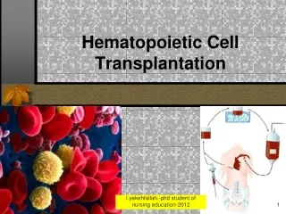
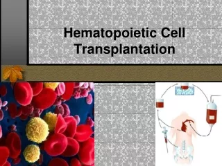
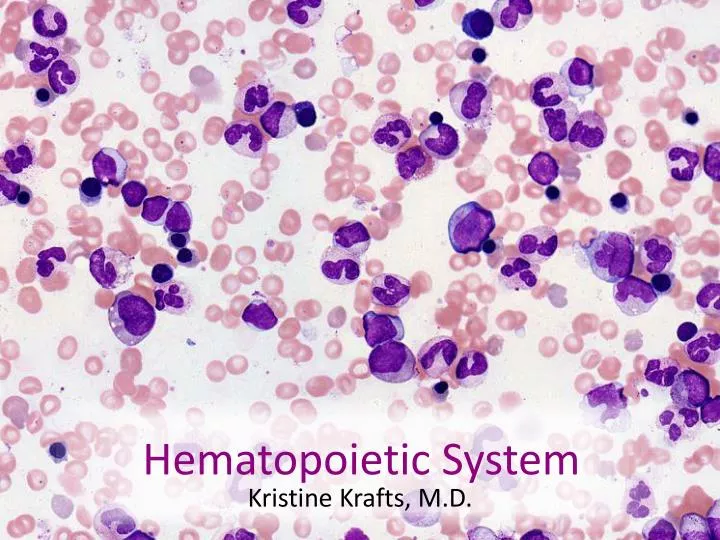
Hematopoietic System. Kristine Krafts, M.D. Hematopoietic System Lecture Objectives. Describe the developmental stages of erythropoiesis. Describe the developmental stages of granulopoiesis. Describe the differences between red marrow and yellow marrow.

E N D
Hematopoietic System Kristine Krafts, M.D.
Hematopoietic System Lecture Objectives • Describe the developmental stages of erythropoiesis. • Describe the developmental stages of granulopoiesis. • Describe the differences between red marrow and yellow marrow. • List the components of bone marrow stroma. • Explain the concept of colony forming units (CFUs).
Hematopoietic System Lecture Outline • Introduction • Erythrocytes • Platelets • Leukocytes • Granulocytes • Agranulocytes
Hematopoietic System Lecture Outline Introduction
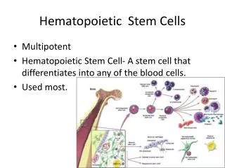
Hematopoietic Tissue Specialized connective tissue. Derived from mesenchyme. Function: produce new blood cells and remove old ones. Two types of hematopoietic tissue • Myeloid tissue (in bone marrow): produces everything except lymphocytes • Lymphoid tissue (in lymph nodes, spleen): produces lymphocytes (different lecture!)
Myeloid Tissue (Bone Marrow) • Bone marrow in adults is found between trabeculae of cancellous (spongy) bone and in the marrow canal of long bones. • Red (hematopoietic) bone marrow is found in flat bones of skull, vertebrae, ribs, pelvis, and ends of long bones. • Yellow bone marrow is found in long bone diaphyses. It is composed of fibrous CT, adipose tissue (yellow color), nerves, and blood vessels.
Components of Bone Marrow • Stroma: a connective tissue network which supports the blood-forming cells. • Sinusoids: wide, thin-walled vessels with discontinuous endothelial cells which allow new blood cells to gain access to the bloodstream. • Developing blood cells: red cell precursors, white cell precursors (except lymphoid precursors), and megakaryocytes.
Stroma Fibers • Type I collagen fibers • Type III collagen (reticular) fibers Cells • Fibroblasts • Macrophages • Adipocytes (fat cells) • Osteogenic cells • Endothelial cells
Bone marrow is present between trabeculae of spongy (cancellous) bones
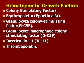
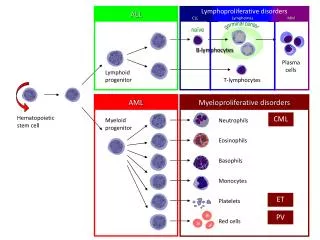
Hematopoiesis • All blood cells arise from a single type of pluripotent stem cell (which is self-renewing). • Pluripotent stem cells divide, forming committed stem cells, which divide and eventually mature into red cells, white cells and platelets. • You need growth factors called “colony stimulating factors” for this process.
Hematopoiesis B-cell T-cell Red cells Platelets Eosinophil Hematopoietic stem cell(self-renewing) Neutrophil Monocyte
Don’t memorize these specific growth factors! Just know they are necessary.
Hematopoietic System Lecture Outline Introduction Erythrocytes
Erythrocyte Maturation • The main jobs: make hemoglobin and form red blood cell with no nucleus. • 2,500,000 red cells released into blood per second! • Start with big cell and big nucleus; size of both decreases as cell matures. • Also: chromatin starts out “fine” (see-through), and becomes progressively more condensed (dark). Nucleus eventually removed.
Morphologic Changes in Erythropoiesis Immature -----------------------------------------------------------------> Mature Small cells, with red (eosinophilic) cytoplasm Large cells, gorgeous basophilic cytoplasm Small, dark blue-black nucleus Large, violet-blue nucleus coarser, then pyknotic nucleus (eventually extruded) Fine chromatin (see nucleoli) Mature red cell Ortho-chromatophilic erythroblast Reticulocyte Polychromatophilic erythroblast Basophilic erythroblast Proerythroblast*
mitotic Proerythroblast* Whole process takes only 7 days! Basophilic erythroblast Polychromatic erythroblast Orthochromatic erythroblast non-mitotic Reticulocyte Mature red cell * Kristine’s favorite cell
Proerythroblast: Big cell, gorgeous sky-blue cytoplasm, very fine chromatin.
Basophilic erythroblast: a little smaller, with more condensed chromatin and darker cytoplasm. Meh.
Polychromatic erythroblasts: even more condensed chromatin, cytoplasm grey-blue (has a little hemoglobin)
Orthochromatic erythroblast: chromatin very condensed, cytoplasm is grey-pink-lavender (has more hemoglobin)
Reticulocytes: no nucleus, cytoplasm is still a little more lavender than mature red cell
Hematopoietic System Lecture Outline Introduction Erythrocytes Platelets
Thrombopoiesis • Platelets come from giant (up to 100 µm) nucleated cells called megakaryocytes. • The megakaryocyte nucleus undergoes endomitosis (replicates DNA without undergoing cell division) • Up to 32 copies of normal DNA! • Multilobed nucleus • Small fragments of megakaryocyte cytoplasm bud off, forming platelets.
Megakaryocyte: huge cell with large, polypoid multilobed nucleus
Hematopoietic System Lecture Outline • Introduction • Erythrocytes • Platelets • Leukocytes • Granulocytes • Agranulocytes
Hematopoietic System Lecture Outline • Introduction • Erythrocytes • Platelets • Leukocytes • Granulocytes
Granulocyte Maturation • 1,250,000 granulocytes (mostly neutrophils) released per second into blood! • Maturation in bone marrow takes about 2 weeks. • Cells spend a few hours in the blood, then leave and stay where needed for a few days, then die.
Morphologic Changes in Granulopoiesis Immature -----------------------------------------------------------------> Mature Cytoplasm loses its basophilic color. Chromatin becomes more condensed. Specific granules increase in number.
Myeloblast mitotic Promyelocyte Myelocyte non-mitotic Metamyelocyte Band cell Segmented neutrophil
Myeloblast: high nuclear:cytoplasmic ratio. Really fine chromatin (nucleoli visible). No granules.
Promyelocyte: biggest cell of the myeloid lineup. Lots of azurophilic (primary) granules.
Myelocyte: has specific granules (and a few azurophilic granules)
Band cell: almost a mature neutrophil – just doesn’t have a segmented nucleus yet.
Hematopoietic System Lecture Outline • Introduction • Erythrocytes • Platelets • Leukocytes • Granulocytes • Agranulocytes
Lymphopoiesis • B lymphocytes develop in the bone marrow. • T lymphocytes develop in the thymus. • B and T cells then leave and populate secondary lymphoid organs (lymph nodes, spleen). • We’ll discuss these later in a different lecture.
Hematopoietic System Lecture Outline • Introduction • Erythrocytes • Platelets • Leukocytes • Granulocytes • Agranulocytes