Download
1 / 56
570 likes | 586 Views
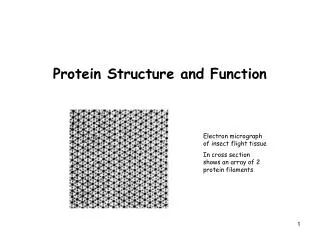
Protein Structure and Function. Proteins. Make up about 15% of the cell Have many functions in the cell Enzymes Structural Transport Motor Storage Signaling Receptors Gene regulation Special functions. Shape = Amino Acid Sequence.

E N D
Proteins • Make up about 15% of the cell • Have many functions in the cell • Enzymes • Structural • Transport • Motor • Storage • Signaling • Receptors • Gene regulation • Special functions
Shape = Amino Acid Sequence • Proteins are made of 20 amino acids linked by peptide bonds • Polypeptide backbone is the repeating sequence of the N-C-C-N-C-C… in the peptide bond • The side chain or R group is not part of the backbone or the peptide bond
Amino AcidsNOTE: You need to know this table Hydrophilic Hydrophobic
Protein Folding • The peptide bond allows for rotation around it and therefore the protein can fold and orient the R groups in favorable positions • Weak non-covalent interactions will hold the protein in its functional shape – these are weak and will take many to hold the shape
Globular Proteins • The side chains will help determine the conformation in an aqueous solution
Hydrogen Bonds in Proteins • H-bonds form between 1) atoms involved in the peptide bond; 2) peptide bond atoms and R groups; 3) R groups
Protein Folding • Proteins shape is determined by the sequence of the amino acids • The final shape is called the conformation and has the lowest free energy possible • Denaturation is the process of unfolding the protein • Can be down with heat, pH or chemical compounds • In the chemical compound, can remove and have the protein renature or refold
Folding@home • The Stanford Folding@home research goal is to understand protein folding, misfolding, and related diseases. • Calculations to create models requires a supercomputer OR many smaller computers (distributed computing). • You can participate by visiting: • Fold@home web site: http://folding.stanford.edu/ • Article on Folding@home: http://www.sciencedaily.com/releases/2002/10/021022070813.htm
Refolding • Molecular chaperones are small proteins that help guide the folding and can help keep the new protein from associating with the wrong partner
Protein Folding • 2 regular folding patterns have been identified – formed between the bonds of the peptide backbone • -helix – protein turns like a spiral – fibrous proteins (hair, nails, horns) • -sheet – protein folds back on itself as in a ribbon –globular protein
Sheets • Core of many proteins is the sheet • Form rigid structures with the H-bond • Can be of 2 types • Anti-parallel – run in an opposite direction of its neighbor (A) • Parallel – run in the same direction with longer looping sections between them (B)
Helix • Formed by a H-bond between every 4th peptide bond – C=O to N-H • Usually in proteins that span a membrane • The helix can either coil to the right or the left • Can also coil around each other – coiled-coil shape – a framework for structural proteins such as nails and skin
Levels of Organization • Primary structure • Amino acid sequence of the protein • Secondary structure • H bonds in the peptide chain backbone • -helix and -sheets • Tertiary structure • Non-covalent interactions between the R groups within the protein • Quanternary structure • Interaction between 2 polypeptide chains
Domains • A domain is a basic structural unit of a protein structure – distinct from those that make up the conformations • Part of protein that can fold into a stable structure independently • Different domains can impart different functions to proteins • Proteins can have one to many domains depending on protein size
Useful Proteins • There are thousands and thousands of different combinations of amino acids that can make up proteins and that would increase if each one had multiple shapes • Proteins usually have only one useful conformation because otherwise it would not be efficient use of the energy available to the system • Natural selection has eliminated proteins that do not perform a specific function in the cell
Protein Families • Have similarities in amino acid sequence and 3-D structure • Have similar functions such as breakdown proteins but do it differently
Proteins – Multiple Peptides • Non-covalent bonds can form interactions between individual polypeptide chains • Binding site – where proteins interact with one another • Subunit – each polypeptide chain of large protein • Dimer – protein made of 2 subunits • Can be same subunit or different subunits
Different Subunit Proteins • Hemoglobin • 2 globin subunits • 2 globin subunits
Protein Assemblies • Proteins can form very large assemblies • Can form long chains if the protein has 2 binding sites – link together as a helix or a ring • Actin fibers in muscles and cytoskeleton – is made from thousands of actin molecules as a helical fiber
Types of Proteins • Globular Proteins – most of what we have dealt with so far • Compact shape like a ball with irregular surfaces • Enzymes are globular • Fibrous Proteins – usually span a long distance in the cell • 3-D structure is usually long and rod shaped
Important Fibrous Proteins • Intermediate filaments of the cytoskeleton • Structural scaffold inside the cell • Keratin in hair, horns and nails • Extracellular matrix • Bind cells together to make tissues • Secreted from cells and assemble in long fibers • Collagen – fiber with a glycine every third amino acid in the protein • Elastin – unstructured fibers that gives tissue an elastic characteristic
Stabilizing Cross-Links • Cross linkages can be between 2 parts of a protein or between 2 subunits • Disulfide bonds (S-S) form between adjacent -SH groups on the amino acid cysteine
Proteins at Work • The conformation of a protein gives it a unique function • To work proteins must interact with other molecules, usually 1 or a few molecules from the thousands to 1 protein • Ligand – the molecule that a protein can bind • Binding site – part of the protein that interacts with the ligand • Consists of a cavity formed by a specific arrangement of amino acids
Formation of Binding Site • The binding site forms when amino acids from within the protein come together in the folding • The remaining sequences may play a role in regulating the protein’s activity
Antibody Family • A family of proteins that can be created to bind to almost any molecule • Antibodies (immunoglobulins) are made in response to a foreign molecule ie. bacteria, virus, pollen… called the antigen • Bind together tightly and therefore inactivates the antigen or marks it for destruction
Antibodies • Y-shaped molecules with 2 binding sites at the upper ends of the Y • The loops of polypeptides on the end of the binding site are what imparts the recognition of the antigen • Changes in the sequence of the loops make the antibody recognize different antigens - specificity
Binding Strength • Can be measured directly • Antibodies and antigens are mixing around in a solution, eventually they will bump into each other in a way that the antigen sticks to the antibody, eventually they will separate due to the motion in the molecules • This process continues until the equilibrium is reached – number sticking is constant and number leaving is constant • This can be determined for any protein and its ligand
Equilibrium Constant • Concentration of antigen, antibody and antigen/antibody complex at equilibrium can be measured – equilibrium constant (K) • Larger the K the tighter the binding or the more non-covalent bonds that hold the 2 together
Enzymes as Catalysts • Enzymes are proteins that bind to their ligand as the 1st step in a process • An enzyme’s ligand is called a substrate • May be 1 or more molecules • Output of the reaction is called the product • Enzymes can repeat these steps many times and rapidly, called catalysts • Many different kinds – see table 5-2, p 168
Enzymes at Work • Lysozyme is an important enzyme that protects us from bacteria by making holes in the bacterial cell wall and causing it to break • Lysozyme adds H2O to the glycosidic bond in the cell wall • Lysozyme holds the polysaccharide in a position that allows the H2O to break the bond – this is the transition state– state between substrate and product • Active site is a special binding site in enzymes where the chemical reaction takes place
Lysozyme • Non-covalent bonds hold the polysaccharide in the active site until the reaction occurs
Enzyme Performance E + S ES EP E + P • Step 1 – binding of the substrate • Limiting step depending on [S] and/or [E] • Vmax – maximum rate of the reaction • Turnover number determines how fast the substrate can be processed = rate of rxn [E] • Step 2 – stabilize the transition state • State of substrate prior to becoming product • Enzymes lowers the energy of transition state and therefore accelerates the reaction
Reaction Rates • KM – [S] that allows rxn to proceed at ½ it maximum rate
Prosthetic Groups • Occasionally the sequence of the protein is not enough for the function of the protein • Some proteins require a non-protein molecule to enhance the performance of the protein • Hemoglobin requires heme (iron containing compound) to carry the O2 • When a prosthetic group is required by an enzyme it is called a co-enzyme • Usually a metal or vitamin • These groups may be covalently or non-covalently linked to the protein
Regulation of Enzymes • Regulation of enzymatic pathways prevent the deletion of substrate • Regulation happens at the level of the enzyme in a pathway • Feedback inhibition is when the end product regulates the enzyme early in the pathway
Feedback Regulation • Negative feedback – pathway is inhibited by accumulation of final product • Positive feedback – a regulatory molecule stimulates the activity of the enzyme, usually between 2 pathways • ADP levels cause the activation of the glycolysis pathway to make more ATP
Allostery • Conformational coupling of 2 widely separated binding sites must be responsible for regulation – active site recognizes substrate and 2nd site recognizes the regulatory molecule • Protein regulated this way undergoes allosteric transition or a conformational change • Protein regulated in this manner is an allosteric protein
Allosteric Regulation • Method of regulation is also used in other proteins besides enzymes • Receptors, structural and motor proteins
Allosteric Regulation • Enzyme is only partially active with sugar only but much more active with sugar and ADP present