Download
1 / 90
940 likes | 1.23k Views
Medical Imaging Ultrasound. Edwin L. Dove 1412 SC edwin-dove@uiowa.edu 335-5635. 3D Reconstruction. Why Ultrasound in Cardiology?. Portable, relatively cheap Non-ionizing During the echocardiogram, it is possible for the cardiologist to: Watch the heart’s motion – in 2D real-time

E N D
Medical ImagingUltrasound Edwin L. Dove 1412 SC edwin-dove@uiowa.edu 335-5635
Why Ultrasound in Cardiology? • Portable, relatively cheap • Non-ionizing • During the echocardiogram, it is possible for the cardiologist to: • Watch the heart’s motion – in 2D real-time • Ascertain if the valves are opening and closing properly, and view any abnormalities • Determine the size of the heart chambers and major vessels • Measure the thickness of the heart walls • Calculate standard metrics of health/disease • e.g., Volume, EF, SV, CO • Dynamic evaluation of abnormalities
p pressure applied in z-direction density viscosity Quantitative Description
Speed of Sound in Tissue • The speed of sound in a human tissue depends on the average density (kg·m3) and the compressibility K (m2·N-1) of the tissue.
Tissue Characteristics • Engineers and scientists working in ultrasound have found that a convenient way of expressing relevant tissue properties is to use characteristic (or acoustic) impedance Z (kg·m-2 ·s-1)
Pressure Generation • Piezoelectric crystal • ‘piezo’ means pressure, so piezoelectric means • pressure generated when electric field is applied • electric energy generated when pressure is applied
Charged Piezoelectric Molecules Highly simplified effect of E field
Reflectance and Refraction Snells’ Law (Assumes i = r)
Reflectivity At normal incidence,i = t = 0 and
Specular Reflection • The first, specular echoes, originate from relatively large, strongly reflective, regularly shaped objects with smooth surfaces. These reflections are angle dependent, and are described by reflectivity equation . This type of reflection is called specular reflection.
Scattered Reflection • The second type of echoes are scattered that originate from small, weakly reflective, irregularly shaped objects, and are less angle-dependent and less intense. The mathematical treatment of non-specular reflection (sometimes called “speckle”) involves the Rayleigh probability density function. This type of reflection, however, sometimes dominates medical images, as you will see in the laboratory demonstrations.
Attenuation • Most engineers and scientists working in the ultrasound characterize attenuation as the “half-value layer,” or the “half-power distance.” These terms refer to the distance that ultrasound will travel in a particular tissue before its amplitude or energy is attenuated to half its original value.
Attenuation • Divergence of the wavefront • Elastic reflection of wave energy • Elastic scattering of wave energy • Absorption of wave energy
Attenuation in Tissue • Ultrasound energy can travel in water 380 cm before its power decreases to half of its original value. Attenuation is greater in soft tissue, and even greater in muscle. Thus, a thick muscled chest wall will offer a significant obstacle to the transmission of ultrasound. Non-muscle tissue such as fat does not attenuate acoustic energy as much. The half-power distance for bone is still less than muscle, which explains why bone is such a barrier to ultrasound. Air and lung tissue have extremely short half-power distances and represent severe obstacles to the transmission of acoustic energy.
Attenuation • As a general rule, the attenuation coefficient is doubled when the frequency is doubled.
Beam Forming • Ultrasound beam can be shaped with lenses • Ultrasound transducers (and other antennae) emit energy in three fields • Near field (Fresnel region) • Focused field • Far field (Fraunhofer region)
A lens will focus the beam to a small spot according to the equation Beam Focusing
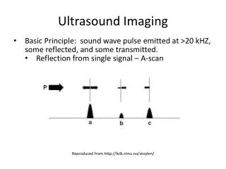
A-mode Ultrasound Amplitude of reflected signal vs. time