Download

1 / 17
170 likes | 217 Views
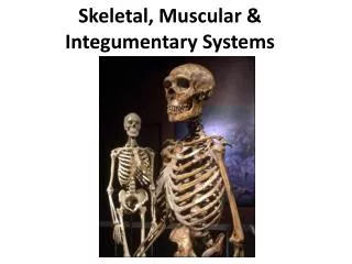
Skeletal Systems. Purpose. Provides form, strength, support and protection for animal’s vital organs (brain, spinal cord, heart, lungs). Layers of Bone. Peridoteum: outermost layer of bone; packed with nerves and blood vessels

E N D
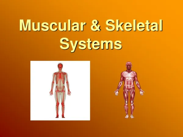
Purpose • Provides form, strength, support and protection for animal’s vital organs (brain, spinal cord, heart, lungs)
Layers of Bone • Peridoteum: outermost layer of bone; packed with nerves and blood vessels • Compact bone: dense, rigid bone under peridoteum; supplies bone with oxygen and nutrients • Bone Marrow: located in central core of bone; produces either white blood cells (to fight infection), red blood cells (for carrying oxygen), or platelets (used to help clot bleeding)
Classes of Bones • 4 classes • 1st class: Long Bones-present in limbs and serve as supporting columns for skeleton and body • 2nd class: Flat Bones-protect the organs and serve as area for muscle attachment • 3rd class: Short Bones-(knee and hock joints)-change direction of tendons • 4th class: Irregular Bones-bones of the vertebrae and cranial base (where is that again??)
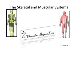
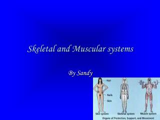
Axial and Appendicular Skeletons • The axial skeleton consists of the skull, vertebral column, and rib cage. • The appendicular skeleton contains the bones of the appendages (limbs, wings, or flippers/fins), and the pectoral and pelvic girdles.
The human skull, or cranium, has a number of individual bones tightly fitted together at immovable joints. At birth many of these joints are not completely sutured together as bone, leading to a number of "soft spots" or fontanels, which do not completely join until the age of 14-18 months.
The vertebral column has 33 individual vertebrae separated from each other by a cartilage disk. These disks allow a certain flexibility to the spinal column, although the disks deteriorate with age, producing back pain. The sternum is connected to all the ribs except the lower pair. Cartilage allows for the flexibility of the rib cage during breathing
The arms and legs are part of the appendicular skeleton. The upper bones of the limbs are single: humerus (arm) and femur (leg). Below a joint (elbow or knee), both limbs have a pair of bones (radius and ulna in the arms; tibia and fibula in legs) that connect to another joint (wrist or ankle). The carpals makeup the wrist joint; the tarsals are in the ankle joint. Each hand or foot ends in 5 digits (fingers or toes) composed of metacarpals (hands) or metatarsals (feet).
Limbs are connected to the rest of the skeleton by collections of bones known as girdles. The pectoral girdle consists of the clavicle (collar bone) and scapula (shoulder blade). • The humerus is joined to the pectoral girdle at a joint and is held in place by muscles and ligaments. A dislocated shoulder occurs when the end of the humerus slips out of the socket of the scapula, stretching ligaments and muscles. The pelvic girdle consists of two hipbones that form a hollow cavity, the pelvis.
The vertebral column attaches to the top of the pelvis; the femur of each leg attaches to the bottom. The pelvic girdle in land animals transfers the weight of the body to the legs and feet. Pelvic girdles in fish, which have their weight supported by water, are primitive; land animals have more developed pelvic girdles. Pelvic girdles in bipeds are recognizable different from those or quadrupeds.
The vertebral column has 33 individual vertebrae separated from each other by a cartilage disk. These disks allow a certain flexibility to the spinal column, although the disks deteriorate with age, producing back pain. The sternum is connected to all the ribs except the lower pair. Cartilage allows for the flexibility of the rib cage during breathing.
Skeletal Disorders • Injury, degenerative wear and tear, and inflammatory disorders affect joints. Sprains are common injuries that cause ligaments to rip of separate from the bone. Tendonitis (such as tennis elbow) and bursitis are inflammations of the tendon sheaths.
Osteoarthritis is a degenerative condition associated with the wearing away of the protective caps of cartilage covering the bone-ends.
Bony growths or spurs develop as the cartilage degenerates, causing restriction of movement and pain. The cause is not known and may just be wear-and-tear associated with aging.
Rheumatoid arthritis is a severely damaging arthritis that begins with inflammation and thickening of the synovial membrane followed by bone degeneration and disfigurement.
More women than men are affected. There may be a genetic predisposition to rheumatoid arthritis. Joint replacement may in some cases restore function.