Download
1 / 51
510 likes | 548 Views
Assessment of the Renal/Urinary System. Anatomy and Physiology Review. Kidneys Ureters Urinary bladder Urethra. Function of the Urinary System. The primary function of the urinary system is to maintain homeostasis Regulate fluids and electrolytes Eliminate waste products Maintain BP

E N D
Anatomy and Physiology Review • Kidneys • Ureters • Urinary bladder • Urethra
Function of the Urinary System The primary function of the urinary system is to maintain homeostasis Regulate fluids and electrolytes Eliminate waste products Maintain BP Involved with RBC production Involved with bone metabolism
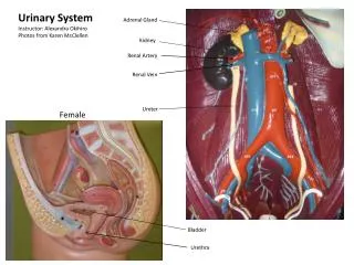
Kidneys Paired Located retroperitoneally on the posterior wall of the abdomen from T12-L3 The average adult kidney weighs 4.5oz The right kidney sits lower in the abdomen due to liver placement An adrenal gland sits on top of each kidney
Kidney Anatomy Each kidney has two parts The renal medulla is the inner portion consists of renal pyramids which are collecting ducts that drain into renal pelvis Once urine leaves the renal pelvis the composition or amount of urine does not change The Cortex is the outer portion contains nephrons
Nephron Each kidney has approximately 1 million nephrons If the function is less than 20% replacement therapy is usually initiated The nephron is responsible for the initial formation of urine
KIDNEY FUNCTIONS Urine formation Excretion of waste products Regulation of electrolytes Regulation of acid-base balance Control of water balance Control BP Regulation of RBC production Synthesis of vitamin D to active form Secretion of prostaglandins Regulation of calcium and phosphorus balance
Urine Formation Urine is formed in the nephrons in a three step process Glomerular filtration Tubular reabsorption Tubular secretion Glomerular Filtration produces ultrafiltrate which enters the tubules Selective reabsorption of H2O & solutes occurs in tubules Selective secretion of solutes occurs in tubules 99% of ultrafiltrate is reabsorbed into the bloodstream 1000-1500mL of urine is produced each day
Excretion of Waste Products The kidney is the body’s main excretory organ The major waste product of protein metabolism is urea 25-30g are produced and excreted daily Other waste products include: Creatinine Phosphates Sulfates Uric acid Drug metabolites
Regulation of Electrolytes In normally functioning kidneys the amount of electrolytes excreted per day is equal to the amount ingested Sodium Linked to blood volume and pressure Significant effects on osmotic pressure 90% of Na in ultrafiltrate is reabsorbed in the proximal tubules and loops of Henle Aldosterone causes kidneys to reabsorb sodium Potassium The kidneys excrete more than 90% of K intake to maintain a normal serum balance Aldosterone causes the kidneys to excrete potassium
Regulation of acid-base balance Normal serum pH is 7.35-7.45 Normal urine pH is 4.6-8 Kidneys 3rd line of defense in acid-base balance respiratory & other buffer systems respond more rapidly kidneys require several hours to a day or more to readjust balance Reabsorb bicarbonate from ultrafiltrate Excrete large quantities of acid in the urine (phosphoric and sulfuric acids) by buffering with ammonia
Control of water balance The human body is made up of 60% water Regulated by Antidiuretic hormone (ADH) or vasopressin Secreted by the posterior pituitary in response to serum osmolality ADH increases reabsorption of water to return serum osmolality to normal Decreased water intake stimulates ADH release ADH controls volume & concentration of urine by regulating permeability of distal tubule to H2O
Control BP The kidney secrets the hormone renin when there is a decrease in BP Renin converts angiotensinogen to angiotensin I Angiotensin I converts to angiotensin II Angiotensin II is a powerful vasoconstrictor and causes BP to increase Increase in BP stops the excretion of renin The adrenal cortex also releases aldosterone in response to increasing serum osmolality or poor perfusion to increase BP
Regulation of RBC production The kidneys release erythropoietin when they sense a decrease in oxygen in the blood Erythropoietin stimulates the bone marrow to produce RBCs
Vitamin D Synthesis The kidneys convert inactive vitamin D to 1,25-dihydroxycholecalciferol Vitamin D is necessary for calcium balance
Ureters • 1 ureter per kidney • Long fibromuscular tubes that connect each kidney to the bladder • Enter bladder at an oblique angle to prevent flow blockage • Propel urine to bladder through peristalsis
Bladder Hollow, muscular organ behind the pubic bone Anatomic capacity is 1500-2000mL Wall of the bladder contains four layers Adventitia—Outer layer/connective tissue Detrusor—smooth muscle Submucosal layer—loose connective tissue Mucosal lining—Inner layer/impermeable to water Bladder neck forms Internal sphincter which is composed of smooth muscle
Urethra Female = 4 cm. (1.5”), Opens anterior to the vagina Male = 20 cm. (8”) 3 sections prostatic = 1”, superior end joins bladder & internal involuntary sphincter; dilatable at this point & larger; has 2 ejaculatory ducts
Urination approximately 150-350 mL of urine triggers stretch receptors & stimulates afferent nerves sending signal to CNS Functional Capacity of bladder is 350mL The efferent pelvic nerve stimulates the bladder to contract, relaxing the urethral sphincter. The decrease in urethral pressure and contractions of the detrusor muscle opens the proximal urethra and leads to flow of urine reflex action however external sphincter can be contracted with voluntary control
TERMINOLOGY Micturation—Urination Frequency—Frequent voiding >every 3hours Urgency—Strong desire to void Hesitancy—Delay or difficulty in initiating voiding Nocturia—Excessive urination at night Oliguria—Output <400mL/day Anuria—Output <50mL/day Polyuria—Increased volume of urine voided Hematuria—RBCs in urine Dysuria—Painful or difficult voiding Enuresis—Involuntary voiding during sleep Incontinence—Involuntary loss of urine
Renal/Urinary System Changes Associated with Aging • Reduced renal blood flow causing kidney loss of cortical tissue by 80 years of age • Thickened glomerular and tubular basement membranes, reducing filtrating ability • Decreased tubule length • Nocturnal polyuria and risk for dehydration • Decreased glomerular filtration rate
Diagnostic Tests Blood Tests Serum Creatinine (0.5 – 1.2 mg/dl) Blood Urea Nitrogen (10-20 mg/dl) BUN/Creatinine Ratio (12:1 to 20:1 mass) Urine Tests Urinalysis Urine for C&S Composite (e.g., 24hr) urine collections Creatinine Clearance Test Urine Electrolytes Osmolality (plasma; urine)
Diagnostic Tests (cont) Bedside sonography; Bladder scanners Radiographic Examinations Kidneys, Ureter, and Bladder X-ray Intravenous Urography Computed Tomography Cystography and Cystourethrography Other Renal Diagnostic Tests Renal Arteriography (Angiography) Renal Biopsy Renography (Kidney Scan) Ultrasonography
Diagnostic Testing: Nursing Implications Anticipate anxiety, discomfort and embarrassment Promote relaxation, comfort and privacy as able Provide teaching
Assessment Techniques • Family history and genetic risk assessment • Demographic data and personal history • Diet history • Socioeconomic status • Current health problems
Physical Assessment • Inspection • Auscultation • Palpation • Percussion • Assessment of the urethra
Blood Tests • Serum creatinine • Blood urea nitrogen • Ratio of blood urea nitrogen to serum creatinine
Urinalysis • Color, odor, and turbidity • Specific gravity • pH • Glucose • Ketone bodies • Protein • Leukoesterase • Cells, casts, crystals, and bacteria
Other Urine Tests • Urine for culture and sensitivity • Composite urine collections • Creatinine clearance—best indication of overall kidney function • Urine electrolytes • Osmolarity, blood/plasma osmolarity, urine osmolarity
Others Diagnostic Tests • Bedside sonography/bladder scanners • Computed tomography • Kidney, ureter, and bladder x-rays • Intravenous urography • Bowel preparation • Allergy information • Fluids
Cystography and Cystourethrography • Instilling dye into bladder via urethral catheter • Voiding cystourethrogram • Monitoring for infection • Encouraging fluid intake • Monitoring for changes in urine output and for development of infection from catheter placement
Renal Arteriography (Angiography) • Possible bowel preparation • Light meal evening before, then nothing by mouth • Injection of radiopaque dye into renal arteries • Assessment for bleeding • Monitoring of vital signs • Absolute bedrest for 4 to 6 hours • Serum creatinine measured for several days to assess effects of test
Renal Biopsy • Percutaneous kidney biopsy • Clotting studies • Preprocedure care • Follow-up care • Assessment for bleeding for 24 hours • Strict bedrest • Monitoring for hematuria • Comfort measures
Renography • Small amount of radioactive material, a radionuclide, used • Procedure via intravenous infection • Follow-up care: • Small amount of radioactive material may be excreted. • Maintain standard precautions. • Client should avoid changing posture rapidly and avoid falling.
Other Diagnostic Tests • Ultrasonography • Cystoscopy and cystourethroscopy • Procedure is invasive. • Consent is required. • Postprocedure care includes monitoring for airway patency, vital signs, and urine output. • Monitor for bleeding and infection. • Encourage client to take oral fluids.
Retrograde Procedures • Retrograde procedures go against the normal flow of urine. • Procedure identifies obstruction or structural abnormalities with the instillation of dye into lower urinary tract. • Monitor for infection. • Follow-up care is the same as for a cystoscopic examination.
Urodynamic Studies • Studies that examine the process of voiding include: • Cystometrography • Urethral pressure profile • Electromyography • Urine stream test
Stress Incontinence Expected Outcomes: to have fewer episodes of stress incontinence or a decreased amount of urine lost with each episode. Interventions: • Nonsurgical • Drug therapy • Behavioral interventions (diet and exercise) • Vaginal Cone therapy • Other: behavior modification, psychotherapy • Surgical • Vaginal, abdominal or retropubic procedures
Urge Urinary Incontinence Expected Outcomes: to use techniques to prevent or manage urge incontinence Interventions: • Drug Therapy (Chart 73-9, p. 1691) • Diet Therapy • Behavioral Interventions • Bladder training • Habit training • Exercise therapy • Electrical stimulation
Reflex Urinary Incontinence Expected outcomes: to achieve continence by keeping urine volume in the bladder within normal limits, preventing bladder overdistention. Interventions: • Drug Therapy • Behavioral Interventions • Bladder Compression (Credé, Valsalva) • Intermittent Self-Cathererization (Clean tech.)