Download
1 / 33
440 likes | 1.7k Views
PROF.T.BAVANISANKAR . CARCINOMA PENIS. Incidence majority – above 60 years less than 40 – 22% less than 30 years – 7% . CHRONIC BALANOPOSTHITIS LEUCOPLAKIA OF GLANS LONGSTANDING GENITAL WARTS

E N D
PROF.T.BAVANISANKAR. CARCINOMA PENIS
Incidence majority – above 60 years less than 40 – 22% less than 30 years – 7%
CHRONIC BALANOPOSTHITIS LEUCOPLAKIA OF GLANS LONGSTANDING GENITAL WARTS PAGET’S DISEASE OF PENIS(ERYTHROPLASIA OF QUERAT) CONDYLOMA ACUMINATA(HPV) BALANITIS XEROTICA OBLITERANS HIV & SEXUALLY TRANSMITTED DISEASES Premalignant lesions
Squamous cell carcinoma Non squamous carcinomas : Basal cell carcinoma Adenocarcinoma Melanoma Sarcoma Metastases Paget’s disease Penile malignancy
INFILTRATING TYPE • PAPILLIFEROUS TYPE • ULCERATIVE TYPE HISTOPATHOLOGY : • Squamous cell ca 59% • Papillary ca 15% • Baseloid 10% • Warty 10% • Verrucous ca 3% • Sarcomatoid 3% PATHOLOGY
Buck’s fascia – temporary natural barrier • Spread of tumour – Local invasion Lymphatic spread Metastasis(liver,lung, bone,brain) Tumour spread
GLANS – 48% PREPUCE – 21% CORONA – 6% SHAFT – 2% SITE OF PRIMARY INVOLVEMENT
EARLY SYMPTOMS – MILD DISCOMFORT, SLIGHT DISCHARGE RECENT ONSET PHIMOSIS LATER – LARGE GROWTH WITH SECONDARY INFECTION – FOUL SMELLING PURULENT AND BLOODY DISCHARGE URETHRAL FISTULA - RARE SIXTY PERCENT – INGUINAL LYMPHNODE ENLARGEMENT – REACTIVE ENLARGEMENT CLINICAL FEATURES
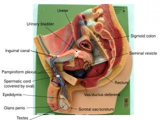
Penile lesion assess site size fixation Involvement of corpora • Bilateral inguinal node examination • Per rectal and bimanual examination for perineal body involvement and presence of pelvic metastasis examination
Prepuce & shaft skin – superficial inguinal nodes • Glans , shaft & corona - superficial inguinal nodes deep inguinal nodes pelvic nodes • Multiple cross-connections at all levels – hence penile lymphatic drainage is bilateral Lymphatic spread
BLOOD INVESTIGATIONS EDGE BIOPSY FNAC OF INGUINAL LYMPH NODES USG – corpora cavernosal involvement MRI-TUMOUR SPREAD INVESTIGATIONS
STAGE I – tumour involving glans or prepuce STAGE II – tumour - body of penis STAGE III – mobile inguinal nodes STAGE IV – adjacent structures or fixed nodes JACKSON’S STAGING
2 cm proximal to the tumor • Urethrostomy is created by approximating the urethra to the surrounding penile skin • Atleast 2cms of shaft should remain • Goal is to preserve • Voiding in standing posture • Sexual function PARTIAL AMPUTATION OF PENIS
TOTAL AMPUTATION OF PENIS TOTAL PENECTOMY A, Incision. B, Transection of the corpora near the level of the pubis. C, Mobilization of the remaining urethra off of the proximal corporal bodies. D, Transposition of the urethra through a curvilinear perineal incision. E, Completion of perinealurethrostomy.
T1 Negative Positive 4 weeks of antibiotics, Observe reassess Negative Positive Follow high risk algorithm
T2-T4, Any T with vascular invasion Any T, Grade III Bilateral Negative Unilateral positive Bilateral positive Mobile < 4cms Mobile < 4cms Bilateral Superficial Inguinal Dissection OR Next slide Next slide Complete Modified Dissection Frozen Section Positive Ipsilateral Deep Inguinal and pelvic node dissection Negative Observe
Unilateral Positive Mobile < 4cms Ipsilateral Inguinal(Superficial+Deep) and pelvic node with Contralateral Superficial Inguinal Dissection Frozen Section Positive Negative Deep Inguinal Observe and pelvic node dissection
Bilateral Positive Mobile < 4cms FNAC Negative Positive Follow bilateral BilateralIlio-inguinal negative algorithm dissection Adjuvant Chemotherapy
Carbon Dioxide (CO2) • Neodymium:yttrium-aluminum-garnet (Nd:YAG) • Potassium titanyl phosphate (KTP) Circumcision is usually recommended at the time of laser surgery if not already done Indications • Carcinoma insitu • T1 lesions • T2 lesions if patient refuses aggressive surgery Laser ablation
Advantages • Good cosmesis • Erectile function not altered in 72% Disadvantages • Exact depth of laser coagulation cannot be assessed • Large lesions cannot be managed • Healing difficulties in obese and immuno-comprised patients Laser ablation
Carcinoma in situ,T1 lesions Advanced inoperable tumour Postoperative RT to inguinal region advanced fixed inguinal nodes Radiotherapy in carcinoma penis
Requisites for Radiotherapy • Circumcision or Dorsal slit – to expose the lesion • Resolution of any surface infection – to prevent maceration & preputial edema Treatment modalities • External beam therapy • Electron beam therapy • Radium mold • Silicon mold • Interstitial therapy(Radium226,Iridium192,Cesium137) Dosage – 5000 to 5700 rads over 3 weeks
Penile necrosis – 10% • Urethral stricture – 30% • Sensory loss • Erectile dysfunction • Testicular damage • Secondary neoplasms Complications of radiotherapy
Bleomycin – radiosensitiser-based regimens 5 FU,METHOTREXATE,BLEOMYCIN,CISPLATIN, VINCRISTINE chemotherapy VBM(Vincristine,Bleomycin,Methotrexate) MBP(Methotrexate,Bleomycin,Cisplatin)