Download
1 / 15
150 likes | 255 Views
Transport of Pharmocokinetic Agents in the Myocardium. Xianfeng Song Sima Setayeshgar Feb. 16, 2004. Pericardial Delivery: Motivation.

E N D
Transport of Pharmocokinetic Agents in the Myocardium Xianfeng Song Sima Setayeshgar Feb. 16, 2004
Pericardial Delivery: Motivation • The pericardial sac is a fluid-filled self-contained space surrounding the heart. As such, it can be potentially used therapeutically as a “drug reservoir.” • Delivery of anti-arrhythmic, gene therapeutic agents to • Coronary vasculature • Myocardium • Recent experimental feasibility • Verrier VL, et al., “Transatrial access to the normal pericardial space: a novel approach for diagnostic sampling, pericardiocentesis and therapeutic interventions,” Circulation (1998) 98:2331-2333. • Stoll HP, et al., “Pharmacokinetic and consistency of pericardial delivery directed to coronary arteries: direct comparison with endoluminal delivery,” Clin Cardiol (1999) 22(Suppl-I): I-10-I-16. Vperi (human) =10ml – 50ml
This work: Outline • Experiments on juvenile farm pigs to measure the spatial concentration profile in the myocardium of agents placed in the pericardial space • Mathematical modeling to investigate the efficacy of agent penetration in myocardial tissue, extract the key physical parameters • Preliminary Results • Conclusions
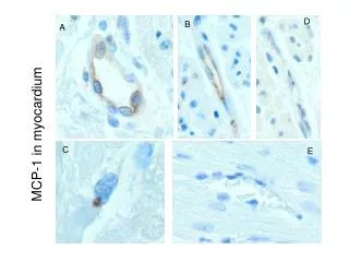
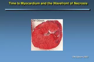
Experiments • Performed by Hans-Peter Stoll, M.D. and Keith L. March, M.D., Ph.D. at Indiana University-Purdue University Indianapolis Medical School • Experimental subjects: juvenile farm pigs • Radiotracer method to determine the spatial concentration profile from gamma radiation rate • Radioiodinated test agents • Insulin-like Growth Factor (125I-IGF, MW: 7740) • Basic Fibroblast Growth Factor (125I-bFGF, MW: 18000)
Mathematical Model • Goals • Investigate the efficacy of agent penetration in myocardium • Extract the key physical parameters • Key physical processes • Substrate transport across boundary layer between pericardial sac and myocardium: a • Substrate diffusion in myocardium: DT • Substrate washout in myocardium (through the vascular and lymphatic capillaries): k
Idealized Spherical Geometry Pericardial sac: R2 – R3 Myocardium: R1 – R2 “Chambers”: 0 – R1 R1 = 2.5cm R2 = 3.5cm Volume of pericardial sac: 10ml-40ml
Governing Equations and Boundary conditions • Governing equation in myocardium CT: concentration of agent in tissue DT: effective diffusion constant in tissue k: washout rate • Consider pericardial sac as a drug reservoir (Well mixing and no washout of drug in pericardial sac) • The drug current flowed through the boundary layer between pericardial sac and myocardium is proportional to the concentration difference between them
Fit to experiments Fitting Error surface
Time-course from simulation Parameters: DT=7×10⁻⁶cm2s-1 k=5×10⁻⁴s-1α=3.2×10⁻⁶cm2s2
Effective Diffusion,D*,in tortuous media • Stokes relation D: diffusion constant R: hydrodynamic radius n: viscosity T: temperature • In tortuous media D*: effective diffusion constant D: diffusion constant in fluid l: tortuosity For myocardium, l = 2.11. (M. Suenson, D.R. Richmond, J.B. Bassingthwaighte, “Diffusion of sucrose, sodium, and water in ventricular myocardium, American Joural of Physiology,” 227(5), 1974 ) • Numerical estimates for diffusion constants • IGF : D ~ • bFGF: D ~ • Our fitted values are in order of 10-6 - 10-5 cm2sec-1 !!
Diffusion in an active viscoelastic medium • Heart tissue is a porous medium consisting of extracellular space and muscle fibers. The extracellular space consists of an incompressible fluid (mostly water) and collagen. • Expansion and contraction of the fiber sheets leads to changes in the gross pore sizes and therefore mixing of the extracellular volume. This effective "stirring" results in larger diffusion constants.
Conclusion • Model is consistent with experiments despite its simplicity. • Numerical determination of values for physical parameters • Effective diffusion constant IGF: D = , bFGF: D = Enhanced diffusion due to motion of heart wall. • Washout rate: k = • Peri-epicardium boundary permeability: a = • Feasibility of computational studies of amount and time course of pericardial drug delivery of drugs to cardiac tissue, using realistic values for physical parameters