Download
1 / 143
1.45k likes | 2.09k Views
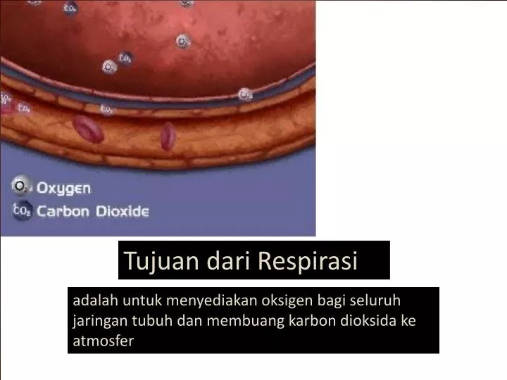
Tujuan dari Respirasi. adalah untuk menyediakan oksigen bagi seluruh jaringan tubuh dan membuang karbon dioksida ke atmosf e r. Hidung. Udara masuk disaring, dihangatkan dan dilembabkan oleh mukosa respirasi. Partikel kasar disaring oleh rambut hidung.

E N D
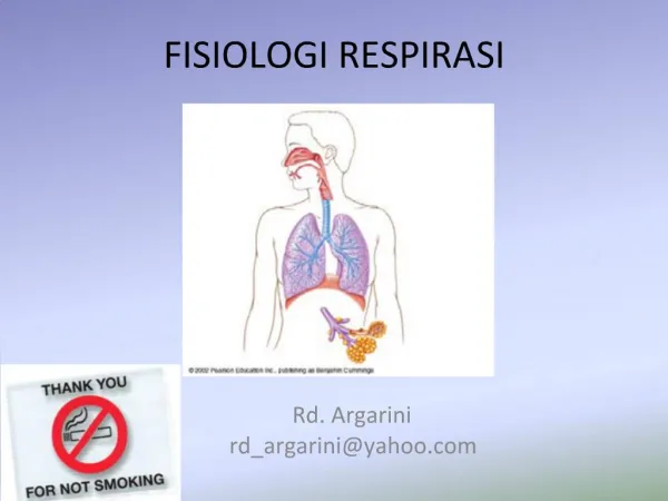
Tujuan dari Respirasi adalahuntukmenyediakanoksigenbagiseluruhjaringantubuhdanmembuangkarbondioksidakeatmosfer
Hidung Udara masuk disaring, dihangatkan dan dilembabkan oleh mukosa respirasi. Partikel kasardisaring oleh rambut hidung. halus: terjerat dalam lapisan mukus. Udara masuk faring: bebas debu, suhu sebanding suhu tubuh, kelembaban hampir 100 %
Rongga torax • Paru adalah organ elastisterletak pada rongga dada/torax. • Paru dilapisi oleh lapisan tipis kontinu yg mengandung kolagen & jar elastis yg disebut PLEURA • Pleura Parietalis melapisi rongga dada sedang Pleura viseralis melapisi paru . • Rongga pleura: ruangan yg memisahkan pleura parietalis & viseralis • Cairan pleura: lapisan tipis antara pleura parietalis dg viseralis berfungsi memudahkan kedua permukaan tersebut bergerak selama pernapasan & untuk mencegah pemisahan torax & paru. • Tekanan rongga pleura < tekanan atmosfer: untuk mencegah kolaps paru.
Rongga torax • 3 faktor yg mempertahankan tekanan negatif intrapleura normal: • Jaringan elastis paru memberikan kekuatan kontinu yg cenderung menarik paru menjauh dr rangka torax. • Kekuatan osmotik yg terdapat di seluruh membran pleura. • Kekuatan pompa limfatik. • Diafragma: otot berbentuk kubah yg membentuk dasar rongga torax & memisahkan rongga tersebut dari rongga abdomen.
Alveoli • Terdapat 2 tipe sel alveolar: • Pneumosit tipe I: lap tipis menyebar & menutupi > 90% daerah permukaan. • Pneumosit tipe II: tanggung jawab pada sekresi surfaktan. • Alveolus: suatu gelembung gas yang dikelilingi oleh jaringan kapiler batas antara cairan & gas membentuk tegangan muka yang cenderung mencegah pengembangan saat inspirasi & cenderung kolaps saat ekspirasi. • Alveolus dilapisi zat lipoprotein (surfaktan) dapat mengurangi tengangan permukaan & resistensi saat inspirasi & mencegah kolaps alveolus (expirasi).
LUNGS 2 Bronchopulmonary segments Lobules Alveolar wall cell types respiratory bronchiole terminal bronchiole pulmonary artery branch simple squamous epithelial cell macrophage (dust cell) septal cell (produces surfactant) alveolar sac Type II alveolar cell making surfactant surfactant Type I alveolar cell macrophage blood capillary respiratory membrane endothelial cell
Alveoli • Pembentukan & pengeluaran surfaktan oleh pneumosit tipe II disintesis secara cepat dari asam lemak yang diekstraksi dari darah, dg kecepatan pergantian yg cepat. Bila aliran darah ke paru terganggu (emboli) akibatnya jumlah surfaktan pada daerah tersebut berkurang. • Produksi surfaktan dirangsang oleh ventilasi aktif, volume tidal yg memadai, hiperventilasi periodik (cepat & dalam) yg dicegah oleh kons O2 yang tinggi (inspirasi). • Pemberian O2 kons tinggi jangka lama (pasien dg ventilasi mekanik) menurunkan produksi surfaktan & menyebabkan kolaps alveolar.
Pernafasan terdiri dari 4 proses : • Ventilasi : Keluar masuknya udara karena adanya selisih tekanan yang terdapat antara atmosfer dan alveolus • Distribusi : Pembagian udara ke cabang -cabang bronkhus • Transportasi dan Difusi - TransportO2 dan CO2 dalam darah dan cairan tubuh ke dandari sel - Difusi O2 dan CO2 antara darah dan alveoliPertukaran gas-gas antara alveoli dan kapiler dipengaruhi oleh tekanan parsial O2 & CO2 dalam atmosfer • Perfusi : Aliran darah yang membawa O2 ke jaringan
JENIS RESPIRASI • RESPIRASI EXTERNAL O2DIBAWA DARI UDARALUARSAMPAIKEKAPILER 2. RESPIRASI INTERNAL O2 DARI KAPILERSAMPAIKESELPADAJARINGAN.
INSPIRATION Active process Boyle’s Law Inspiratory muscles Phrenic nerves (C3-5) Thoracic nerves (T1 – T11) Process thoracic volume pleural volume intrapleural pressure lung volume intrapulmonic pressure air flow into the lungs 760 mmHg 760 mmHg 760 mmHg 758 mmHg FORCED INSPIRATION BEFORE INSPIRATION DURING INSPIRATION
EXPIRATION Passive process at rest elastic recoil surface tension internal intercostals (11 pairs) Process thoracic volume pleural volume intrapleural pressure lung volume intrapulmonic pressure air flow of the lungs external abdominal oblique internal abdominal oblique transversus abdominis rectus abdominis Forced expiration 760 mmHg internal intercostals (11 pairs) rectus abdominis abdominal obliques transversus abdominis 762 mmHg
COMPLIANCE Compliance is the ease with which the lungs and thoracic wall can be expanded during inspiration. Related to two factors: elasticity surface tension Compliance is decreased with any condition that: destroys lung tissue (emphysema) fills lungs with fluid (pneumonia) produces surfactant deficiency (premature birth, near-drowning) interferes with lung expansion (pneumothorax)
PULMONARY VOLUMES, CAPACITIES, AND RATES maximum inspiration Volumes 6000 ml tidal volume (500 ml) 5000 ml IRV anatomical dead space (150 ml) alveolar ventilation (350 ml) physiological dead space VC TLC 4000 ml inspiratory reserve volume (3000 ml) expiratory reserve volume (1200 ml) residual volume (1300 ml) 3000 ml TV Capacities 2000 ml ERV total lung capacity (TV+IRV+ERV+RV) vital capacity (TV+IRV+ERV) (4700 ml) inspiratory capacity (TV+IRV) functional residual capacity (RV+ERV) 1000 ml RV maximum expiration SPIROGRAM Rates maximum voluntary ventilation = TV x breaths/minute alveolar ventilation rate = alveolar ventilation x breaths/minute
Kontrol Pernapasan • Otot pernapasan diatur oleh neuron & reseptor pada pons & medula oblongata. • Faktor utama pengaturan pernapasan: respon dari pusat kemoreseptor dalam pusat pernapasan terhadap tekanan persial CO2 dan pH darah arteri
Kontrol Pernapasan • Persarafan parasimpatis/ kolinergik (mll nervus fagus) menyebabkan kontraksi otot polos bronkus shg menyebabkan bronkokonstriksi & peningkatan sekresi kel mukosa & sel goblet. • Rangsangan simpatis ditimbulkan epinefrin mll reseptor adrenergik-beta2 menyebabkan relaksasi otot polos bronkus, bronkodilatasi, & berkurangnya sekresi bronkus. • Sistem saraf nonkolinergik non adrenergik (NANC): melibatkan berbagai mediator seperti ATP, oksida nitrat, substance P, dan VIP (vasoactive intestinal peptide) respon penghambatan, meliputi bronkodilatasi, dan diduga berfungsi sebagai penyeimbang terhadap fungsi pemicuan oleh sistem kolinergik.
Signs and Symptoms of Pulmonary Disease • Dyspnea – subjective sensation of uncomfortable breathing, feeling “short of breath” • Ranges from mild discomfort after exertion to extreme difficulty breathing at rest. • Usually caused by diffuse and extensive rather than focal pulmonary disease.
Dyspnea cont. • Due to: • Airway obstruction • Greater force needed to provide adequate ventilation • Wheezing sound due to air being forced through airways narrowed due to constriction or fluid accumulation • Decreased compliance of lung tissue
Signs of dyspnea: • Flaring nostrils • Use of accessory muscles in breathing • Retraction (pulling back) of intercostal spaces
BATUK • Batukmerupakangejalaterseringpenyakitpernapasan • Batuk merupakan reflex pertahanan yang timbul akibat iritasi percabangan trakeobronkial • Batuk yang berlangsung lebih dari 3 minggu harus diselidiki untuk memastikan penyebabnya. • Bronkhitis kronik, asma, tubercolosis dan pneomonia merupakan penyakit yang secara tipikal memiliki batuk sebagai gejala yang mencolok
Cough may result from: • Inflammation of lung tissue • Increased secretion in response to mucosal irritation • Inhalation of irritants • Intrinsic source of mucosal disruption – such as tumor invasion of bronchial wall • Excessive blood hydrostatic pressure in pulmonary capillaries • Pulmonary edema – excess fluid passes into airways
When cough can raise fluid into pharynx, the cough is described as a productive cough, and the fluid is sputum. • Production of bloody sputum is called hemoptysis • Usually involves only a small amount of blood loss • Not threatening, but can indicate a serious pulmonary disease • Tuberculosis, lung abscess, cancer, pulmonary infarction.
Cough that does not produce sputum is called a dry, nonproductive or hacking cough. • Acute cough is one that resolves in 2-3 weeks from onset of illness or treatment of underlying condition. • Us. caused by URT infections, allergic rhinitis, acute bronchitis, pneumonia, congestive heart failure, pulmonary embolus, or aspiration.
A chronic cough is one that persists for more than 3 weeks. • In nonsmokers, almost always due to postnasal drainage syndrome, asthma, or gastroesophageal reflux disease • In smokers, chronic bronchitis is the most common cause, although lung cancer should be considered.
SPUTUM • Pembentukan sputum: Orangdewasa normal mukussekitar 100 ml dalamsalurannapastiaphariMukusdiangkutmenuju faring dengangerakanpembersihansilia yang melapisisaluranpernapasanbilamukusberlebihanprosespembersihantidakefektif mukustertimbunmembranmukosaakanterangsangmukusdibatukkankeluarsebagai sputum.
SPUTUM • Sputum yang berwarna kekuning-kuningan menunjukkan infeksi. • Sputum yang berwarna hijau merupakan petunjuk penimbunan nanahtimbul karena adanya verdoperoksidase yang dihasilkan oleh polimorfonuklear (PMN). • Sputum yang berwarna merah muda dan berbusa merupakan tanda edema paru akut. • Sputum yang berlendir lekat dan warna abu-abu atau putih merupakan tanda bronkhitis kronik. Sedangkan sputum yang berbau busuk merupakan tanda abses paru atau bronkiektasis.
Cyanosis • When blood contains a large amount of unoxygenated hemoglobin, it has a dark red-blue color which gives skin a characteristic bluish appearance. • Most cases arise as a result of peripheral vasoconstriction – result is reduced blood flow, which allows hemoglobin to give up more of its oxygen to tissues- peripheral cyanosis. • Best seen in nail beds • Due to cold environment, anxiety, etc.
Central cyanosis can be due to : • Abnormalities of the respiratory membrane • Mismatch between air flow and blood flow • Expressed as a ratio of change in ventilation (V) to perfusion (Q) : V/Q ratio • Pulmonary thromboembolus - reduced blood flow • Airway obstruction – reduced ventilation • In persons with dark skin can be seen in the whites of the eyes and mucous membranes.
Jari Tabuh Jaritabuhadalahperubahanbentuk normal falang distal dan kuku tangandan kaki sertaditandaidengan Kehilangansudut kuku yang normalnya 160 derajat. Rasa halusberonggapadadasar kuku. Ujung jarimenjadibesar. jaritabuhberhubungandenganpeyakitparu (TB, absesparu, ataukankerparu). Penyakitkardiovaskuler (tetralogifallotatauendokarditisinfektif) ataupenyakithatikronik
Next… 2. Hipoksia (O2 yang tidak adekuat dalam tingkat jaringan) dan Hipoksemia (PaO2 dibawah normal normal 80-100 mmhg). Tanda dan gejala hipoksemia dan hipoksia tidak spesifik dan mencakup takipnea, dispnea, sakit kepala, pikiran yang bingung, takikardi, dan sianosis. 3. Hipokapnia dan hiperkapnia Hipokapnia didefinisikan sebagai menurunnya PaCO2 <35 mmhg. Penyebab langsung selalu hiperventilasi alveolar (eliminasi CO2 lebih cepat daripada produksinya). Hiperkapnia / asidosis respiratorius merupakan meningkatnya PaCO2 >45 mmhg. Penyebab langsung adalah selalu hipoventilasi alveolar (kegagalan dalam mengeliminasi CO2 secepat produksinya).
Pain • Originates in pleurae, airways or chest wall • Inflammation of the parietal pleura causes sharp or stabbing pain when pleura stretches during inspiration • Usually localized to an area of the chest wall, where a pleural friction rub can be heard • Laughing or coughing makes pain worse • Common with pulmonary infarction due to embolism
Inflammation of trachea or bronchi produce a central chest pain that is pronounced after coughing • Must be differentiated from cardiac pain • High blood pressure in the pulmonary circulation can cause pain during exercise that often mistaken for cardiac pain (angina pectoris)
Respiratory Disorders • Respiratory disorder can be classified into different group • Respiratory tract infection Common cold,Influenza,Pneumonias,T.B • Disorder of lung inflation Pleural pain and pleural effusion • Obstructive air way disorders Bronchial asthma, COPD, Emphysema, Bronchitis • Pulmonary vascular disorder • Lung cancer
INFLUENZA Penyakit yang disebabkanoleh virus influenza. Gejala yang ditimbulkanantara lain pilek, hidungtersumbat, bersin- bersin, dantenggorokanterasagatal. Perludiketahui virus iniselaluhanyabisamenembussaluranpernafasanatassaja , sehinggabisadisimpulkansaluranrespirasi yang lebihdalamsangatresistenimmunterhadap virus ini.
Asthma is a chronic inflammatory disorder of the airways in whichmany cells and cellular elements play a role In susceptible individuals, this inflammation causes recurrent episodes of wheezing, breathlessness, chest tightness and coughing, particularly at night or in the early morning..
Causes and Triggers • Allergies such as to pollens, mold spores, pet dander, and dust mites • Infections (colds, viruses, flu, sinus infection) • Exercise • Aspirin or nonsteroidal anti-inflammatory drug (NSAID) hypersensitivity, sulfite sensitivity • Use of beta-adrenergic receptor blockers (including ophthalmic preparations)
Cont…… • Irritants such as strong odors from perfumes or cleaning solutions, air pollution, and tobacco smoke • Weather (changes in temperature and/or humidity, cold air) • Strong emotions such as anxiety, laughter, crying, and stress • Industrial triggers (wood, grain dust, cotton dust, isocyanate containing paints, aluminum, hair spray, penicillins) • Beta blockers even in form of eye drops
Gastroesophageal reflux disease • Chronic sinusitis or rhinitis • OSA (obstructive sleep apnoe) • Obesity • Alergy bronchopulmonary aspergilosis Comorbid condition