Download
1 / 4
40 likes | 134 Views
Morphologic appearance of regression in thin cutaneous melanomas.

E N D
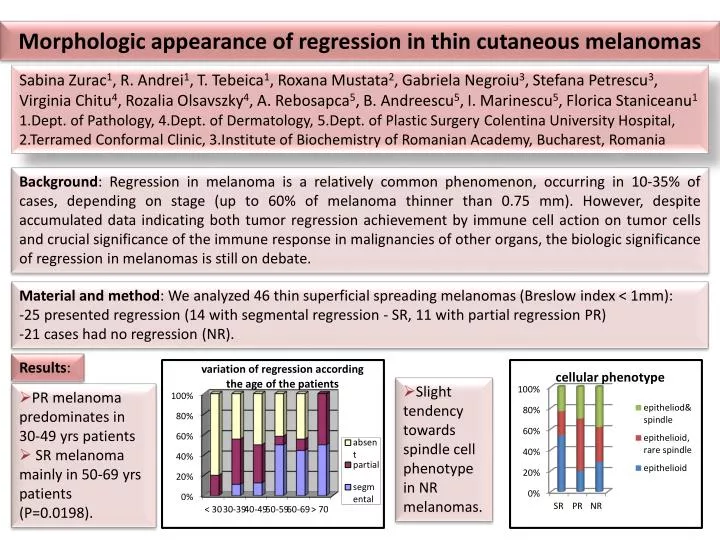
Morphologic appearance of regression in thin cutaneous melanomas Sabina Zurac1, R. Andrei1, T. Tebeica1, Roxana Mustata2, Gabriela Negroiu3, Stefana Petrescu3, Virginia Chitu4, RozaliaOlsavszky4, A. Rebosapca5, B. Andreescu5, I. Marinescu5, FloricaStaniceanu1 1.Dept. of Pathology, 4.Dept. of Dermatology, 5.Dept. of Plastic Surgery Colentina University Hospital, 2.Terramed Conformal Clinic, 3.Institute of Biochemistry of Romanian Academy, Bucharest, Romania Background: Regression in melanoma is a relatively common phenomenon, occurring in 10-35% of cases, depending on stage (up to 60% of melanoma thinner than 0.75 mm). However, despite accumulated data indicating both tumor regression achievement by immune cell action on tumor cells and crucial significance of the immune response in malignancies of other organs, the biologic significance of regression in melanomas is still on debate. • Material and method: We analyzed 46 thin superficial spreading melanomas (Breslow index < 1mm): • 25 presented regression (14 with segmental regression - SR, 11 with partial regression PR) • 21 cases had no regression (NR). Results: • Slight tendency towards spindle cell phenotype in NR melanomas. • PR melanoma predominates in 30-49 yrs patients • SR melanoma mainly in 50-69 yrs patients (P=0.0198).
Regression absent in melanomas with high mitotic index (P=0.029) with tendency towards fewer mitoses in SR than PR and/or NR melanomas. • SR melanomas tend to have larger regression areas with lesser fibroplasia than PR cases. Partial regression in melanoma with Breslow 0.22. • Very thin melanomas (Breslow <0.45 mm) tends to associate PR. Partial regression in melanoma without noticeable mitoses. Partial regression with moderate fibrosis in the regressed area.
Tumor infiltrating lymphocytes (TIL) are almost significantly more numerous in SR melanomas than NR ones (P=0.06), while PR and NR melanomas have similar TIL distribution. • The more extended, the thicker were the areas of regression in SR melanomas. Plasma cells in segmental regression • SR melanomas associate presence of plasma cells within tumor inflammatory infiltrate (P=0.0173), while PR melanomas do not (P=0.21). Relatively frequent TILs in melanoma with segmental regression in other areas
Conclusions: In thin melanomas, partial and segmental regression seem to belong to different spectrum of alteration. SR melanomas present features usually associated with better prognosis (older age, low mitotic index, TIL presence) There are significantly more numerous plasma cells within SR areas, suggesting an antibodies-mediated immunological mechanism rather than a cellular one as previously expected. Extended area of segmental regression Acknowledgments: project partially supported by Postdoctoral Program POSDRU/89/1.5/S/60746