Download
1 / 127
1.27k likes | 1.28k Views
Arne Elofsson. MicroArrays and proteomics. Introduction. Microarrays Introduction Data threatment Analysis Proteomics Introduction and methodologis Data threatment Analysis The network view of biology Connectivity vs function. Topics. Goal – study many genes at once

E N D
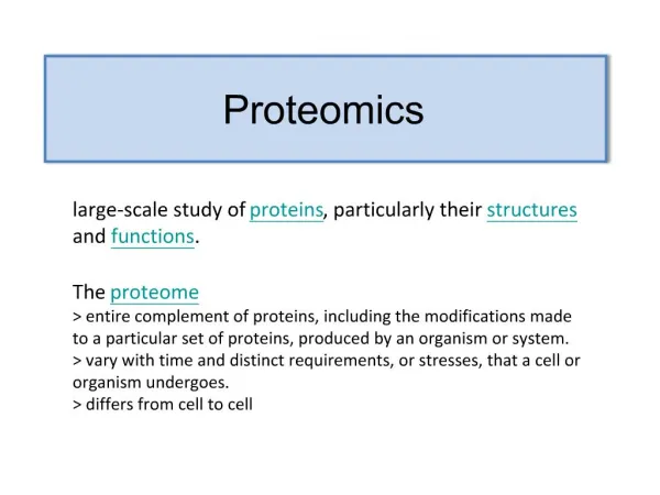
Arne Elofsson MicroArrays and proteomics
Introduction • Microarrays • Introduction • Data threatment • Analysis • Proteomics • Introduction and methodologis • Data threatment • Analysis • The network view of biology • Connectivity vs function
Topics • Goal – study many genes at once • Major types of DNA microarray • How to roll your own • Designing the right experiment • Many pretty spots – Now what? • Interpreting the data
The Goal “Big Picture” biology – • What are all the components & processes taking place in a cell? • How do these components & processes interact to sustain life? One approach: What happens to the entire cell when one particular gene/process is perturbed?
Genome Sequence Flood • Typical results from initial analysis of a new genome by the best computational methods: For 1/3 of the genes we have a “good”idea what they are doing (high similarity to exp. studied genes) For 1/3 of the genes, we have a guess at what they are doing (some similarity to previously seen genes) For 1/3 of genes, we have no idea what they are doing (no similarity to studied genes)
Large Scale Approaches • Geneticists used to study only one (or a few) genes at a time • Now, thousands of identified genes to assign biological function to • Microarrays allow massively parallel measurements in one experiment (3 orders of magnitude or greater)
Southern and Northern Blots • Basic DNA detection technique that has been used for over 30 years: • Northern Blots • Hybridizing labelled DNA to a solid support with RNA from cells. • Southern blots: • A “known” strand of DNA is deposited on a solid support (i.e. nitrocellulose paper) • An “unknown” mixed bag of DNA is labelled (radioactive or fluorescent) • “Unknown” DNA solution allowed to mix with known DNA (attached to nitro paper), then excess solution washed off • If a copy of “known” DNA occurs in “unknown” sample, it will stick (hybridize), and labeled DNA will be detected on photographic film
The process Building the chip: MASSIVE PCR PCR PURIFICATION AND PREPARATION PREPARING SLIDES PRINTING RNA preparation: Hybridizing the chip: POST PROCESSING CELL CULTURE AND HARVEST ARRAY HYBRIDIZATION RNA ISOLATION cDNA PRODUCTION DATA ANALYSIS PROBE LABELING
The arrayer Ngai Lab arrayer , UC Berkeley Print-tip head
Pins collect cDNA from wells 384 well plate Contains cDNA probes Print-tip group 1 cDNA clones Spotted in duplicate Print-tip group 6 Glass Slide Array of bound cDNA probes 4x4 blocks = 16 print-tip groups
Image Duplicate spots Scanning Detector PMT
Microarray summary Create 2 ssamples Label one green and one red Mix in equal amounts and hybridze in array Process images and normalize data Read data
Microarray life cyle Biological Question Data Analysis & Modelling Sample Preparation MicroarrayDetection Taken from Schena & Davis Microarray Reaction
Biological question Differentially expressed genes Sample class prediction etc. Experimental design Microarray experiment 16-bit TIFF files Image analysis (Rfg, Rbg), (Gfg, Gbg) Normalization R, G Estimation Testing Clustering Discrimination Biological verification and interpretation
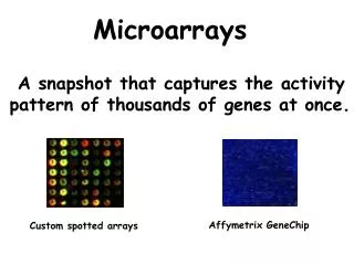
Non gene expression arrays CHIP-CHIP ARRAYS immunoprecipitation to micro-arrays that contain genomic regions (ChIP-chip) has provided investigators with the ability to identify, in a high-throughput manner, promoters directly bound by specific transcription factors. SNPs Genomic (tiling) arrays Different types of Arrays • Gene Expression arrays • cDNA (Brown/Botstein) • One cDNA on each spot • Spotted • Affymetrix • Short oligonucleotides • Photolithography • Ink-jet microarrays from Agilent • 25-60-mers “printed directly on glass slides • Flexible, rapid, but expensive
Affymetrix Gene Chips expensive ($500 or more) limited types avail, no chance of specialized chips fewer repeated experiments usually more uniform DNA features Can buy off the shelf Dynamic range may be slightly better Pros/Cons of Different Technologies Spotted Arrays • relative cheap to make (~$10 slide) • flexible - spot anything you want • Cheap so can repeat experiments many times • highly variable spot deposition • usually have to make your own • Accuracy at extremes in range may be less
Data processing • Image analysis • Normalisation • Log2 transformation
Cy5 Cy3 Image Analysis & Data Visualization Cy5 Cy3 log2 Cy3 Cy5 Experiments 8 4 2 fold 2 4 8 Underexpressed Overexpressed Genes
Why Normalization ? To remove systematic biases, which include, • Sample preparation • Variability in hybridization • Spatial effects • Scanner settings • Experimenter bias
What Normalization Is & What It Isn’t • Methods and Algorithms • Applied after some Image Analysis • Applied before subsequent Data Analysis • Allows comparison of experiments • Not a cure for poor data.
Sample Preparation Scanning + Image Analysis Hybridization Normalization Data Analysis Array Fabrication Where Normalization Fits In Normalization Subsequent analysis, e.g clustering, uncovering genetic networks Spot location, assignment of intensities, background correction etc.
Choice of Probe Set • House keeping genes – e.g. Actin, GAPDH • Larger subsets – Rank invariant sets Schadt et al (2001) J. Cellular Biochemistry37 • Spiked in Controls • Chip wide normalization – all spots Normalization method intricately linked to choice of probes used to perform normalization
Form of Data Working with logged values gives symmetric distribution Global factors such as total mRNA loading and effect of PMT settings easily eliminated.
Mean & Median Centering • Simplistic Normalization Procedure • Assume No overall change in D.E. Mean log (mRNA ratio) is same between experiments. • Spot intensity ratios not perfect log(ratio) log(ratio) – mean(log ratio) or log(ratio) log(ratio) – median(log ratio) more robust
0 0 Location & Scale Transformations Mean & Median centering are examples of location transformations
Regression Methods • Compare two hybridizations (exp. and ref) – use scatter plot • If perfect comparability – straight line through 0, slope 1 • Normalization – fit straight line and adjust to 0 intercept and slope 1 • Various robust procedures exist
log R M A log G M = log R – log G A = ½[ log R + log G ] M = Minus A = Add M-A Plots M-A plot is 45° rotation of standard scatter plot 45°
M M A A M-A Plots Un-normalized Normalized Normalized M values are just heights between spots and the “general trend” (red line)
Methods To Determine General Trend • Lowess (loess) Y.H. Yang et al, Nucl. Acid. Res. 30 (2002) • Local Average • Global Non-linear Parametric Fit e.g. Polynomials • Standard Orthogonal decompositions e.g. Fourier Transforms • Non-orthogonal decompositions e.g. Wavelets
Lowess Gasch et al. (2000) Mol. Biol. Cell 11, 4241-4257
M A Lowess Demo 1
M A Lowess Demo 2
M A Lowess Demo 3
M A Lowess Demo 4
M A Lowess Demo 5
M A Lowess Demo 6
M A Lowess Demo 7
Things You Can Do With Lowess (and other methods) Bias from different sources can be corrected sometimes by using independent variable. • Correct bias in MA plot for each print-tip • Correct bias in MA plot for each sector • Correct bias due to spatial position on chip
Pros & Cons of Lowess • No assumption of mathematical form – flexible • Easy to use • Slow - unless equivalent kernel pre-calculated • Too flexible ? Parametric forms just as good and faster to fit.
What is BASE? • BioArray Software Environment • A complete microarray database system • Array printing LIMS • Sample preparation LIMS • Data warehousing • Data filtering and analysis
What is BASE? • Written by Carl Troein et al at Lund University, Sweden • Webserver interface, using free (open source and no-cost) software • Linux, Apache, PHP, MySQL
Why use BASE? • Intergrated system for microarray data storage and analysis • MAGE-ML data output • Sharing of data • Free • Regular updates/bug fixes
Features of BASE • Password protected • Individual / group / world access to data • New analysis tools via plugins • User-defined data output formats
Using BASE • Annotation • Array printing LIMS • Biomaterials • Hybridization • Analysis