Download
1 / 1
10 likes | 207 Views
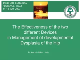
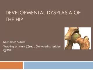
FOLLOW-UP OF THE ORTHOPAEDIC TREATMENT OF DEVELOPMENTAL DYSPLASIA OF THE HIP BY SONOGRAPHY. Azzoni Roberto, Cabitza Paolo, Parrini Matteo Orthopaedic Dept., State University of Milan – 20097 San Donato Milanese (Milan) 30, via Morandi, Italy (E-mail: roberto.azzoni@unimi.it).

E N D
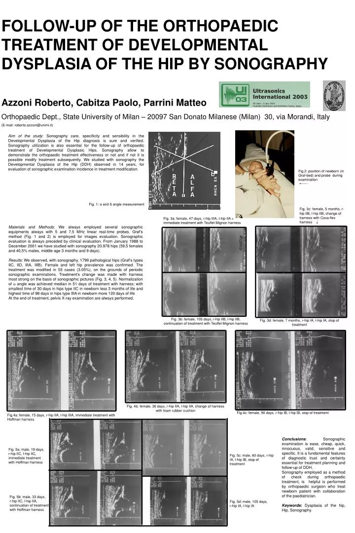
FOLLOW-UP OF THE ORTHOPAEDIC TREATMENT OF DEVELOPMENTAL DYSPLASIA OF THE HIP BY SONOGRAPHY Azzoni Roberto, Cabitza Paolo, Parrini Matteo Orthopaedic Dept., State University of Milan – 20097 San Donato Milanese (Milan) 30, via Morandi, Italy (E-mail: roberto.azzoni@unimi.it) Aim of the study: Sonography care, specificity and sensibility in the Developmental Dysplasia of the Hip diagnosis is sure and verified. Sonography utilization is also essential for the follow-up of orthopaedic treatment of Developmental Dysplasic Hips. Sonography allow to demonstrate the orthopaedic treatment effectiveness or not and if not it is possible modify treatment subsequently. We studied with sonography the Developmental Dysplasia of the Hip (DDH) observed in 14 years, for evaluation of sonographic examination incidence in treatment modification. Fig.2: position of newborn (in Graf-bed) and probe during examination Fig. 1: α and ß angle measurement Fig. 3c:female, 5 months, r-hip IIB, l-hip IIB, change of harness with Coxa-flex harness Fig. 3a: female, 47 days, r-hip IIIA, l-hip IIA > immediate treatment with Teuffel-Mignon harness Materials and Methods: We always employed several sonographic equipments always with 5 and 7,5 MHz linear real-time probes. Graf’s method (Fig. 1 and 2) is employed for images evaluation. Sonographic evaluation is always preceded by clinical evaluation. From January 1988 to December 2001 we have studied with sonography 20.978 hips (59,5 females and 40,5% males, middle age 3 months and 9 days). Results: We observed, with sonography, 1799 pathological hips (Graf’s types IIC, IID, IIIA, IIIB). Female and left hip prevalence was confirmed. The treatment was modified in 55 cases (3.05%), on the grounds of periodic sonographic examinations. Treatment’s change was made with harness most strong on the basis of sonographic pictures (Fig. 3, 4, 5). Normalization of angle was achieved median in 51 days of treatment with harness; with smallest time of 30 days in hips type IIC in newborn less 3 months of life and highest time of 98 days in hips type IIIA in newborn more 120 days of life At the end of treatment, pelvis X-ray examination are always performed. Fig. 3b: female, 105 days, r-hip IIB, l-hip IIB, continuation of treatment with Teuffel-Mignon harness Fig. 3d: female, 7 months, r-hip IA, l-hip IA, stop of treatment Fig. 4b: female, 36 days, r-hip IIA, l-hip IIA, change of harness with foam rubber cushion Fig.4c: female, 90 days, r-hip IB, l-hip IB, stop of treatment Fig.4a: female, 15 days, r-hip IIA, l-hip IIIA, immediate treatment with Hoffman harness Conclusions: Sonographic examination is ease, cheap, quick, innocuous, valid, sensitive and specific. It is a fundamental features of diagnostic trust and certainty essential for treatment planning and follow-up of DDH. Sonography employed as a method of check during orthopaedic treatment, is helpful is performed by orthopaedic surgeon who treat newborn patient with collaboration of the paediatrician. Keywords: Dysplasia of the hip, Hip, Sonography. Fig. 5a: male, 19 days, r-hip IIC, l-hip IIC, immediate treatment with Hoffman harness Fig. 5c: male, 60 days, r-hip IA, l-hip IB, stop of treatment Fig. 5b: male, 33 days, r-hip IIC, l-hip IIA, continuation of treatment with Hoffman harness Fig. 5d: male, 105 days, r-hip IA, l-hip IA