Download
1 / 32
320 likes | 340 Views
Discover various pathologies observable in EKG readings, including arrhythmias, myocardial infarctions, pericarditis, and chamber hypertrophy. Learn about ECG electrodes, waveforms, and interpreting heart activity.

E N D
What types of pathology can we identify and study from EKGs? • Arrhythmias • Myocardial ischemia and infarction • Pericarditis • Chamber hypertrophy • Electrolyte disturbances (i.e. hyperkalemia, hypokalemia) • Drug toxicity (i.e. digoxin and drugs which prolong the QT interval)
PR QRS AH HV How does the heart work
AV node activated by Atrial depolarizationSends signal through His-purkinje bundleGet depolarization of SEPTUM Left and Right BUNDLES transmit signal to Left and Right VENTRICLESNet “Vector” towards the LVShould be narrow (<120msec) if bundles working properlyThen have REPOLARIZATION = TwaveThe appearance of this electrical activity depends on which lead you are using to look at it
How to Look at an ECG • Rate: Is the heart rate too fast or slow? • Rhythm: Sinus rhythm or not? • Axis: Where does the majority of electrical activity point? • P wave: How big are the atria? • PR interval: How healthy is the AV node? • QRS wave: Is there abnormal conduction or a ventricular source? • QT: Long is bad • Ischemia and hypertrophy
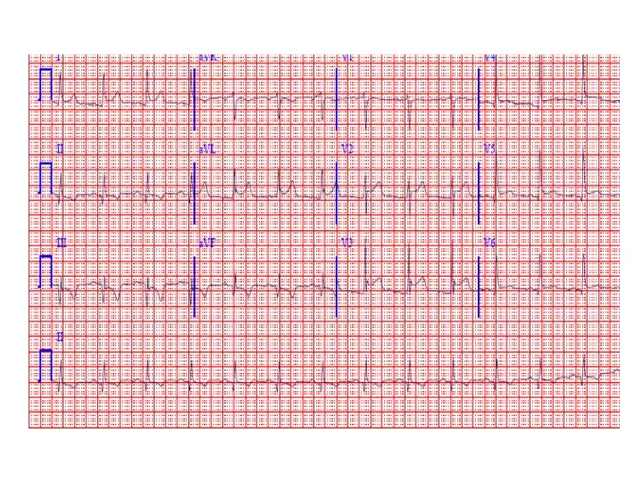
ECG PaperCan Determine Heart Rate Rule: 300, 150, 100, 75, 60, 50 counting over for each big sqaure
What is the heart rate? Answer = 75 per min
Rhythm : Is there a p wave? = Sinus Is it followed by a QRS?
Irregular pacemaker Multifocal atrial rhythm Atrial fibrillation Atrial fib/flutter Ectopic beats PVC PAC PJC Irregular conduction AV node block 1st degree: PR interval > 200 msec 2nd degree: Type 1: Wenkebach Type 2: dropped beat 3rd degree: p waves marching independent to QRS Reasons to have an irregular rhythm
Examples of Rhythms Multifocal Atrial Rhythm AFIB Atrial Flutter AFIB V TACH
EKG Leads 3 Standard Limb Leads 3 Augmented Limb Leads 6 Precordial Leads The standard EKG has 12 leads: The axis of a particular lead represents the viewpoint from which it looks at the heart.
The QRS QRS < 120 msec QRS > 120 msec Rabbit ears in V1 & V2 Wide S wave in V5 & V6 R axis deviation QRS > 120 msec Deep slurred S wave in V1 Wide R wave in V6, I & avL L axis deviation
Review • Rate: Is the heart rate too fast or slow? • Rhythm: Sinus rhythm or not? • Axis: Where does the majority of electrical activity point? • P wave: How big are the atria? • PR interval: How healthy is the AV node? • QRS wave: Is there abnormal conduction or a ventricular source? • QT: Long is bad • Ischemia and hypertrophy