Download
1 / 73
740 likes | 955 Views
Stem Cells in the Vascular System. Kristina Boström May 2005. Stem Cell - Definition. Cells that undergo asymmetric division resulting in self-renewal of the parent stem cell as well as a daughter cell capable of differentiating down specific lineages.

E N D
Stem Cells in the Vascular System Kristina Boström May 2005
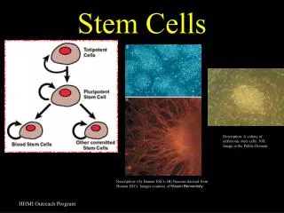
Stem Cell - Definition Cells that undergo asymmetric division resulting in self-renewal of the parent stem cell as well as a daughter cell capable of differentiating down specific lineages. The tissue specific, committed stem cells provide a supply of terminally differentiated cells for physiologic tissue turnover for the life of the individual.
Stem Cell Plasticity A stem cell, which is committed to give rise to the expected tissues, may differentiate into cells other than these expected tissues, the so-called unexpected tissues. Termed TRANSDIFFERENTIATION. However, it is equally plausible that uncommitted stem cells exist within the tissue and have the potential of differentiating into many or all tissues. It is hard to generate convincing data (Wagers & Weissman, Cell 2004). Committed stem cell Uncommitted stem cell Horwitz. Arch Med Res, 2004
Endothelial Progenitor Cells (EPC) Mesenchymal Stem Cells (MSC)
Layers of the Vascular Wall Endothelium Internal elastic lamina Media External elastic lamina Adventitia Differentiation between the arterial and venous side of the vasculature.
Normal and Diseased Vessel Wall Hillebrands et al. ATVB 2003
“The Vascular Tree” Tree-like three-dimensional structure with branches and branch points.
Formation of Endothelial Tubes VASCULOGENESIS ANGIOGENESIS Drake. Birth Defects Res 2003 Goumans et al. Trends Cardiovasc Med 2003
Embryonic Vasculogenesis Drake. Birth Defects Res 2003
Adult Vasculogenesis Iwami et al. J Cell Mol Med 2004
Areas of Potential Adult Vasculogenesis Atherosclerotic plaques Tumor formation Bone disorders Inflammatory diseases Drake. Birth Defects Res 2003
ARTERIOGENESIS Recruitment of SMC precursors and SMC differentiation Stenmark & Abman. Annu Rev Physiol 2005
The tenet of stem cell biology is that the cells only differentiate into cell types associated with the tissue from which they were isolated. Called into question! - more plastic than thought.
Hemangioblasts - Cells with hematopoietic and endothelial potential Were believed to exists only in embryos Endothelial progenitor cells (EPC) can be isolated from peripheral blood mononuclear cells (PBMNC) by flow cytometry by e.g. CD34 which previously was associated with hematopoietic stem cells. Overturned Dogma!!
ENDOTHELIAL PROGENITOR CELLS (EPC) • 0.0001 - 0.02% of peripheral blood cells • CD34+ AC133+ VEGFR2+ lin- cells • Study vasculogenesis • Determine progenitor state in patients • Clinical trials of expanded cell populations
Asahara et al. Science 1997 Isolated putative ECP from human peripheral blood. Two antigens shared by angioblasts and HSC: CD34 and Flk-1 (= VEGFR-2 and KDR). Tested for incorporation of EPC in three animal models Human MBC34+ cells into athymic mice with hindlimb ischemia- (heterologous transplantation). ß-Gal overexpressing mice MB, MBFlk1+ or MBFlk1- injected into mice on same background but without ß-Gal. Incorporation in hind limb ischemia. - (homologous transplantation). Injected DiI-labeled rabbit CD34+ or CD34- MB into rabbits with hindlimb ischemia. Found DiI labeling 1-6 weeks afterwards in ischemic limb. - (autologous transplantation).
EPC Isolated from Human Blood Asahara et al. Science 1997;275:964
Incorportion on EPC from Peripheral Blood into Ischemic Hindlimb Autologous Rabbit Model Asahara et al. Science 1997;275:964
Evidence of a CD34+ cells from BM and circulation differentiate into EPC Grown in presence of bFGF, IGF-1 and VEGF Stained positive for CD34 and VEGFR2. Stained for vWF and took up Ac-LDL. Tested in Canine BM transplant model with genetically distinct donor and recipient. 6-8 months after BM transplant, Dacron graft impervious to in-growth of vessels was implanted in the descending aorta. 12 weeks later, only donor cells covered the Dacron graft. Shi et al. Blood 1998;92:362
J. Clin. Invest. 2002;109:337 Multipotent adults progenitor cells (MAPC) from human BM Murine Lewis lung carcinoma spheroids in NOD-SCID mice Studied tumor angiogenesis
Anti Mouse CD31 Anti Human ß2-microglobulin Anti- vWF Reyes et al. J. Clin. Invest. 2002;109:337
(Jiang et al. PNAS 2004) To determine the EC potential of human BM and PBC, blood vessels in sex-matched transplant recipients were evaluated None of the >4,000 ECs examined had more than two sex chromosomes, consistent with an absence of cell fusion. Y chromosome signals were not detected in sex-matched female recipients, excluding the vertical transmission of male cells. None of the recipients evaluated before hematopoietic engraftment demonstrated donor-derived ECs, indicating a close linkage between the recovery of hematopoiesis and EC outcomes.
BM-Derived Endothelial Cells Jiang et al. PNAS 2004
Putative Cascade and Expressional Profiles of Human BM-derived EPC Differentiation Iwami et al. J Cell Mol Med 2004
Endothelial Progenitor Cells Positive for CD34 and VEGFR2 expression. Sometimes CD133 (AC133, prominin) - more likely to reflect immature progenitor cells. CD34+/VEGFR2+ cells may also represent shedded EC of the vessel wall. Proof of EC characteristics after outgrowth and differentiation in vitro. May also be isolated from fetal liver or umbilical cord blood. No data on lifetime in vivo of EPC under physiological or pathological conditions.
EPC - From Bone Marrow to Vasculature Iwami et al. J Cell Mol Med 2004
Important factors for mobilization and proliferation of EPC: • Physiological • Age • Gender (estrogen) • Embryonal development • Exercise • Pathologic • Smoking • Stable coronary artery disease • Myocardial infarction (tissue ischemia) • Vascular trauma • Drugs • Statins • Growth factors • VEGF • G-CSF/GM-CSF • SDF-1 • Erythropoietin • PPARgamma
Growth factors may Enhance population Compensate for decline in population Explain aging in EPC
EPC dependent on what environment it enters into. Poor endothelialization usually leads to enhanced vascular disease. Diabetes mellitus: decreased proliferation capacity, reduced adhesiveness and ability to form capillary tubes in vitro. Diabetics shed more EC into circulation. Hypercholesterolemia - dysfunction in mature EC.
Chemotaxis, Adhesion, Migration SDF-1 attracts progenitors to ischemic tissues CXCR4 VEGF b2-integrins and a4b1-integrins are capable of mediating cell-cell interactions important for adhesion.
Differentiation of EPC Regulation largely unknown, but the entire VEGF response system is critical
Role in Physiology vs Pathology Therapeutic Potential Urbich & Dimmeler Circ Res 2004
Neovascularization • Circulating mature EC do not improve • neovascularization. • Tissue injury stimulate EPC incorporation. • Incorporation varies in the literature, between 0>50%. • The >50% predominantly detected in models of tumor • angiogenesis. • Even if low incorporation, EPC may have other • characteristics that promote neovascularization such as • release of proangiogenic factors.
High recruitment of BM-derived EPC into Growing Tumors Lyden et al. Nat. Med. 2001
Endothelial Regeneration • Dacron vascular grafts and ventricular assist devices • covered by endothelial progenitors. • Denudation of artery after balloon injury re-endothelialized. • Rapid re-endothelialization may improve atherosclerosis • and prevent restenosis.
EPC Contribute to Re-Endothelialization after Vascular Injury Carotid Injury Model EC visualized using FITC-labeled lection Werner et al. Circ. Res. 2003
Therapeutic Applications of EPC Iwami et al. J Cell Mol Med 2004
Potential for therapy of EPC Critical limb ischemia Myocardial infarction Vascular grafts Stroke Pulmonary hypertension Diabetic retinopathy Neoplasm
Necessity to develop standardized methods to isolates, phenotype, and evaluate quality of cells. The number in the circulation may limit therapeutic use.
Layers of the Vascular Wall Endothelium Internal elastic lamina Media External elastic lamina Adventitia Differentiation between the arterial and venous side of the vasculature.
The bone marrow contains two apparently discrete populations of stem cells. In addition to the HSC/EPC, there are also bone marrow stromal cells or mesenchymal stem cells (MSC). Less characterized than the HSC/EPC and its exact location within the bone marrow is less clear. Low density in bone marrow aspirate.
Markers for MSC Adherent cells WGA binding and Sca-1 (null mice, late osteoporosis) Enriched population: Sca-1+Lin-CD31-CD45- 30% plating efficiency STRO-1+ : Includes all CFU-F CD105 (endoglin) Negative for CD34, CD45, CD11b
Location of MSC in bone marrow Most likely in the vessel wall in the bone marrow. Would be similar to vascular smooth muscle cells and pericytes, or endosteal cells. Cultured MSC Express alpha-SMC (70%) H-caldesmon Metavinculin Calponin SM-MHC Proteins constituting basal lamina Similar response to PDGF as pericytes STRO-1+ Potential for differentiation into a variety of cell types
Bone Marrow Pericytes