Download
1 / 24
250 likes | 504 Views
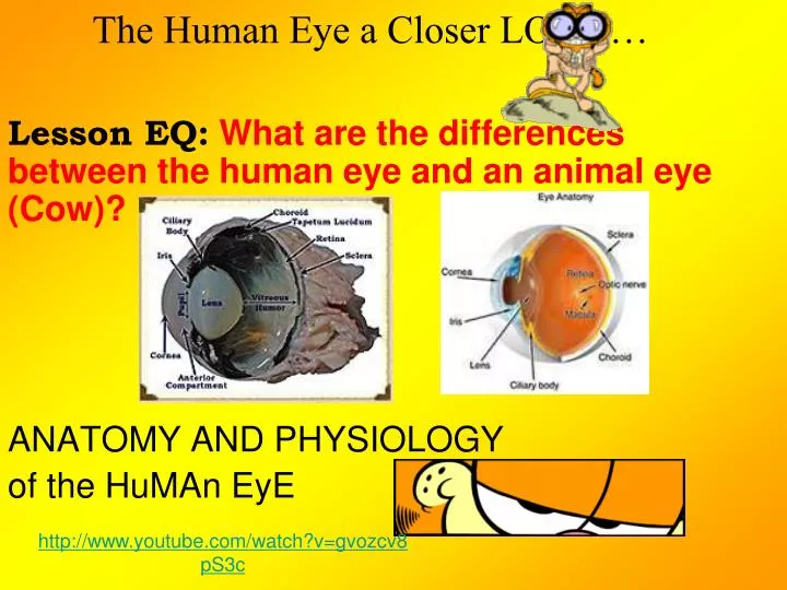
The Human Eye a Closer LOOK…. Lesson EQ: What are the differences between the human eye and an animal eye (Cow)? ANATOMY AND PHYSIOLOGY of the HuMAn EyE. http://www.youtube.com/watch?v=gvozcv8pS3c. HuMan EyE Anatomy & Physiology.

E N D
The Human Eye a Closer LOOK… Lesson EQ:What are the differences between the human eye and an animal eye (Cow)? ANATOMY AND PHYSIOLOGY of the HuMAn EyE http://www.youtube.com/watch?v=gvozcv8pS3c
HuMan EyE Anatomy & Physiology • Aqueous Humor: Clear, watery fluid found in the anterior chamber of the eye. • Choroid: Layer of blood vessels that nourish the eye; also, because of the high melanocytes content, the choroid acts as a light-absorbing layer. • Cornea: Transparent tissue covering the front of the eye. Does not have any blood vessels; does have nerves. • Iris: Circular band of muscles that controls the size of the pupil. The pigmentation of the iris gives "color" to the eye. Blue eyes have the least amount of pigment; brown eyes have the most. • Lens: Transparent tissue that bends light passing through the eye. To focus light, the lens can change shape by bending. • Pupil: Hole in the center of the eye where light passes through. • Retina: Layer of tissue on the back portion of the eye that contains cells responsive to light (photoreceptors). • Rods: Photoreceptors responsive in low light conditions. • Cones: Photoreceptors responsive to color and in bright conditions. • Sclera: Protect coating around the posterior five-sixths of the eyeball. • Vitreous Humor: Clear, jelly-like fluid found in the back portion of the eye. Maintains shape of the eye.
HuMaNeYe Anatomy and physiology • http://www.kscience.co.uk/animations/eye.swf
COW vs HUMAN EyES • The cow's eye is bigger, its iris is only one color. • The cow has a tapetum-what allows animals to see well in the dark • The iris is brown on a cow and on humans its many different colors. • Cow eyes are on the side of their heads offering a wider field of view to watch for predators, our eyes are in the front to be a better predator.
Now that we have learned some anatomy of the eye and physiology, lets look at why vision can be distorted and corrected!
BLAME IT On the Choroid! What causes "red eye" when you take a flash photograph? • The choroid is a layer of tissue at the back of the eye that contains a large number of blood vessels. "Red eye" usually happens when a flash photograph is taken in dim light. In dim light, the pupil is dilated and allows plenty of light to enter the eye. "Red eye" is caused when the choroid reflects the light of the flash. The pupil does not constrict fast enough to reduce the entering light and the flash light reflects back out of the eye and is recorded on film. Some cameras use red eye reduction methods: a short burst of light is emitted before the film is exposed. The brief burst of light allows the pupil to constrict and thus reduces "red eye.
COLOR BLINDNESS • Color blindness is the inability to differentiate between different colors. The most common type is red-green color blindness. This occurs in 8 percent of males and 0.4 percent of females. It occurs when either the red or green cones are not present or not functioning properly. People with this problem are not completely unable to see red or green, but often confuse the two colors. This is an inherited disorder and affects men more commonly since the capacity for color vision is located on the X chromosome. (Women have two X chromosomes, so the probability of inheriting at least one X with normal color vision is high; men have only one X chromosome to work with.
THE CARROT MYTH When severe vitamin A deficiency is present, then night blindness occurs. Vitamin A is necessary to form retinal, which is part of the rhodopsin molecule. Vitamin A deficiency can cause night blindness, says John Allred, a professor of nutrition at Ohio State University. An extreme deficiency can even cause blindness. Vitamin A deficiency is the leading cause of blindness in the Third World. But if you're not deficient in vitamin A, your vision won't improve no matter how many carrots or other beta-carotene-rich fruits and vegetables you eat. When the levels of light-sensitive molecules are low due to vitamin A deficiency, there may not be enough light at night to permit vision. During daylight, there is enough light stimulation to produce vision despite low levels of retinal.
Visual Acuity • Vision or visual acuity is tested by reading a Snellen eye chart at a distance of 20 feet. • If you have 20/20 vision, it means that when you stand 20 feet away from the chart you can see what a "normal" human being can see. (In metric, the standard is 6 meters and it's called 6/6 vision). • In other words, if you have 20/20 vision your vision is "normal" -- a majority of people in the population can see what you can see at 20 feet. If you have 20/40 vision, it means that when you stand 20 feet away from the chart you can only see what a normal human can see when standing 40 feet from the chart. • 20/200 is the cutoff for legal blindness in the United States. • You can also have vision that is better than the norm. A person with 20/10 vision can see at 20 feet what a normal person can see when standing 10 feet away from the chart. • Hawks, owls and other birds of prey have much more acute vision than humans. A hawk has a much smaller eye than a human being but has lots of sensors (cones) packed into that space. This gives a hawk vision that is eight times more acute than a human's. A hawk might have 20/2 vision!
ERRORS OF REFRACTION • Normally, your eye can focus an image exactly on the retina: • Nearsightedness and farsightedness occur when the focusing is not perfect. • When nearsightedness (myopia) is present, a person is able to see near objects well and has difficulty seeing objects that are far away. Light rays become focused in front of the retina. This is caused by an eyeball that is too long, or a lens system that has too much power to focus. Nearsightedness is corrected with a concave lens. This lens causes the light to diverge slightly before it reaches the eye, as seen below: When farsightedness (hyperopia) is present, a person is able to see distant objects well and has difficulty seeing objects that are near. Light rays become focused behind the retina. This is caused by an eyeball that is too short, or by a lens system that has too little focusing power. This is corrected with a convex lens, as seen above:
Astigmatism and Age • Astigmatism is an uneven curvature of the cornea and causes a distortion in vision. To correct this, a lens is shaped to correct the unevenness. Why does vision worsen as we age? • As we grow older, the lens becomes less elastic. It loses its ability to change shape. This is called presbyopia and is more noticeable when we try to see things that are close up, because the ciliary body must contract to make the lens thicker. The loss of elasticity prevents the lens from becoming thicker. As a result, we lose the ability to focus on close objects. • To correct this, bifocals are required. Bifocals are a combination of a lower lens for close vision (reading) and an upper lens for distance vision.
Conditions that can lead to blindness: • Legal blindness is usually defined as visual acuity less than 20/200 with corrective lenses. • Cataracts - This is a cloudiness in the lens that blocks light from reaching the retina. It becomes more common as we age, but babies can be born with a cataract. As it worsens, it can require surgery to remove the lens and place an intraocular lens. • Glaucoma - If the aqueous humor does not drain out correctly, then pressure builds up in the eye. This causes the cells and nerve fibers in the back of the eye to die. This can be treated with medications and surgery. • Diabetic retinopathy - Persons with diabetes can get blockage of blood vessels, leakage of blood vessels and scarring that can lead to blindness. This can be treated with laser surgery. • Trauma - Direct trauma or chemical injuries can cause enough damage to the eyes to prevent adequate vision. • There are many other causes of blindness, such as vitamin A deficiency, tumors, strokes, neurological diseases, other infections, hereditary diseases and toxins.
Thanks for hanging around!!!!Time for a QUIZ!!!! • http://www.childrensuniversity.manchester.ac.uk/interactives/science/brainandsenses/eye.asp
AFTER IMAGE • An afterimage or ghost image or image burn-in is an optical illusion that refers to an image continuing to appear after the exposure to the original image has ceased. • Stare at the next pictures for 60 seconds then quickly look at a blank white computer paper.