Download
1 / 20

E N D
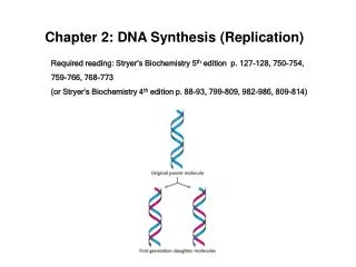
1. Chapter 3: DNA Replication Linnea Fletcher Ph.D.
BIOL 2316
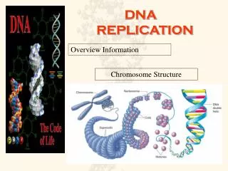
2. Principal Points Explain DNA replication
Precursors, enzymes, direction etc in E. coli
Compare and contrast the DNA pol in prokaryotes to eukaryotes.
When does DNA replication occur in prokaryotes? In eukaryotes?
Explain how telomerase works to maintain the length of linear chromosomes
4. Figure 3.1 Three models for DNA replication. (a) Semiconservative model (the correct model). (b) Conservative model. (c) Dispersive model. The parental strands are shown in taupe, and the newly synthesized strands are shown in red. Figure 3.1 Three models for DNA replication. (a) Semiconservative model (the correct model). (b) Conservative model. (c) Dispersive model. The parental strands are shown in taupe, and the newly synthesized strands are shown in red.
5. How did Meselson & Stahl�s experiment show that cells replicate by semiconservative replication?
How could this experiment be used to show how an alien replicates DNA? (you might want to know this)
6. Figure 3.1 Schematic diagram for separating DNAs of different buoyant densities by equilibrium centrifugation in a cesium chloride density gradient. The separation of 14N-DNA and 15N-DNA is illustrated. Figure 3.1 Schematic diagram for separating DNAs of different buoyant densities by equilibrium centrifugation in a cesium chloride density gradient. The separation of 14N-DNA and 15N-DNA is illustrated.
7. Figure 3.2 The Meselson�Stahl experiment. The demonstration of semiconservative replication in E. coli. Cells were grown in a 15N-containing medium for several replication cycles and then were transferred to a 14N-containing medium. At various times over several replication cycles, samples were taken; the DNA was extracted and analyzed by CsCl equilibrium density gradient centrifugation. Shown in the figure are a schematic interpretation of the DNA composition after various replication cycles, photographs of the DNA bands, and densitometric scans of the bands. Figure 3.2 The Meselson�Stahl experiment. The demonstration of semiconservative replication in E. coli. Cells were grown in a 15N-containing medium for several replication cycles and then were transferred to a 14N-containing medium. At various times over several replication cycles, samples were taken; the DNA was extracted and analyzed by CsCl equilibrium density gradient centrifugation. Shown in the figure are a schematic interpretation of the DNA composition after various replication cycles, photographs of the DNA bands, and densitometric scans of the bands.
8. What are the reaction components necessary for in vitro synthesis of DNA? Describe how they interact in the mechanism of DNA polymerization.
What direction does is the new strand of DNA grown by DNA polymerases?
What have the 5 DNA polymerases discovered in E. coli been called, and what are their biological roles in the cell?
What is the importance of the 5�?3� exonuclease activity of DNA polymerases I and III?
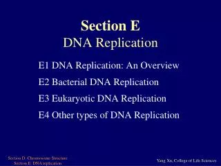
9. Discuss the process of replication of DNA in prokaryotes, explaining the roles played by the following:
Opening up the DNA double helix to form a replication bubble with two replication forks:
origins of replication,
ii. the initiator protein,
the DNA helicase enzyme
topoisomerases
the single-strand binding protein
building a primer with the primase enzyme,
assembling the complementary strands
the DNA polymerase III enzyme,
the nucleotide triphosphates,
the leading strand, the lagging strand, & the Okazaki fragments
DNA gyrase, a topoisomerase
Removing the primer with DNA polymerase I
Joining the Okazaki fragments with DNA ligase
10. Figure 3.4 DNA chain elongation catalyzed by DNA polymerase. (a) Mechanism at molecular level. (b) The same mechanism, using a shorthand method to represent DNA. Figure 3.4 DNA chain elongation catalyzed by DNA polymerase. (a) Mechanism at molecular level. (b) The same mechanism, using a shorthand method to represent DNA.
12. Figure 3.6 Model for the events occurring around a single replication fork of the E. coli chromosome. (a) Initiation. (b) Further untwisting and elongation of the new DNA strands. (c) Further untwisting and continued DNA synthesis. (d) Removal of the primer by DNA polymerase I. (e) Joining of adjacent DNA fragments by the action of DNA ligase. Green=RNA; red=new DNA.Figure 3.6 Model for the events occurring around a single replication fork of the E. coli chromosome. (a) Initiation. (b) Further untwisting and elongation of the new DNA strands. (c) Further untwisting and continued DNA synthesis. (d) Removal of the primer by DNA polymerase I. (e) Joining of adjacent DNA fragments by the action of DNA ligase. Green=RNA; red=new DNA.
13. the nick. the nick.
16. Figure 3.13 Temporal ordering of DNA replication initiation events in replication units of eukaryotic chromosomes. Figure 3.13 Temporal ordering of DNA replication initiation events in replication units of eukaryotic chromosomes.
17. Why is telomerase classified as a reverse transcriptase enzyme?
What types of cell lines require telomerase activity?
Why don�t normal (nontransformed) cells just get more and more repeats of DNA at the end of their chromosomes as a result of telomerase activity?
18. Figure 3.14 The problem of replicating completely a linear chromosome in eukaryotes. (a) Schematic diagram of a parent doublestranded DNA molecule representing the full length of a chromosome. (b) After semiconservative replication, new DNA segments hydrogen bonded to the template strands have RNA primers at their ends. (c) The RNA primers are removed, DNA polymerase fills the resulting gaps, and DNA ligase joins the adjacent fragments. However, at the two telomeres, there are still gaps at the ends of the new DNA. The gaps result from RNA primer removal, because no new DNA synthesis could fill them in. Figure 3.14 The problem of replicating completely a linear chromosome in eukaryotes. (a) Schematic diagram of a parent doublestranded DNA molecule representing the full length of a chromosome. (b) After semiconservative replication, new DNA segments hydrogen bonded to the template strands have RNA primers at their ends. (c) The RNA primers are removed, DNA polymerase fills the resulting gaps, and DNA ligase joins the adjacent fragments. However, at the two telomeres, there are still gaps at the ends of the new DNA. The gaps result from RNA primer removal, because no new DNA synthesis could fill them in.
19. Figure 3.15 Synthesis of telomeric DNA by telomerase. The example is of Tetrahymena telomeres. The process is described in the text. (a) The starting point is the chromosome end with5� gap left after primer removal. (b) Binding of telomerase to the overhanging telomere repeat at the end of the chromosome. (c) Synthesis of three-nucleotide DNA segment at chromosome end, using the RNA template of telomerase. (d) The telomerase moves so that the RNA template can bind to the newly synthesized TTG in a different way. (e) Telomerase catalyzes the synthesis of a new telomere repeat, using the RNA template. The process recurs, to add more telomere repeats. (f) After telomerase has left, new DNA is made on the template, starting with an RNA primer. (g) After the primer is removed, the result is a longer chromosome than at the start, with a new 5� gap. Figure 3.15 Synthesis of telomeric DNA by telomerase. The example is of Tetrahymena telomeres. The process is described in the text. (a) The starting point is the chromosome end with5� gap left after primer removal. (b) Binding of telomerase to the overhanging telomere repeat at the end of the chromosome. (c) Synthesis of three-nucleotide DNA segment at chromosome end, using the RNA template of telomerase. (d) The telomerase moves so that the RNA template can bind to the newly synthesized TTG in a different way. (e) Telomerase catalyzes the synthesis of a new telomere repeat, using the RNA template. The process recurs, to add more telomere repeats. (f) After telomerase has left, new DNA is made on the template, starting with an RNA primer. (g) After the primer is removed, the result is a longer chromosome than at the start, with a new 5� gap.
20. Figure 3.16 Assembly of new nucleosomes at a replication fork. New nucleosomes are assembled first with the use of either a parental or a new H3�H4 tetramer and then by completing the structure with a pair of H2A�H2B dimers. Figure 3.16 Assembly of new nucleosomes at a replication fork. New nucleosomes are assembled first with the use of either a parental or a new H3�H4 tetramer and then by completing the structure with a pair of H2A�H2B dimers.