Download
1 / 33
350 likes | 426 Views
Brain tumors. Incidence of tumors. 10-25 per 100.000 population per year 5-15% among all tumors. Classification of brain tumors. Tumors of neuroepithelial tissue Astrocytic tumors Oligodendroglial tumors Ependymal tumors Mixed gliomas Choroid plexus tumors

E N D
Incidence of tumors • 10-25 per 100.000 population per year • 5-15% among all tumors
Classification of brain tumors • Tumors of neuroepithelial tissue • Astrocytic tumors • Oligodendroglial tumors • Ependymal tumors • Mixed gliomas • Choroid plexus tumors • Neuronal and mixed neuronal-glial tumors • Pineal parenchymal tumors • Embrional tumors
Classification of brain tumors • Tumors of cranial and spinal nerves • Tumors of the meninges • Tumors of meningothelial cells • Mesenchymal nonmeningothelial tumors • Primary melanocytic lesions • Tumors of sellar region • Germ cell tumors • Lymphomas and hemopoietic neoplasms • Cysts and tumor-like lesions • Local extentions from regional tumors • Metastatic tumors • Unclassified tumors
Surgical classification of brain tumors • Intracerebral • Intraventricular • Extracerebral • Supratentorial • Infratentorial • Exact localization (lobes, basal ganglia, brain stem, ventricles)
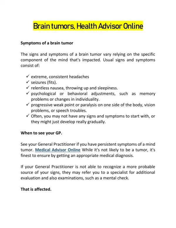
Clinical presentations • Symptoms due to increased intracranial pressure • Headache • Vomiting • Seizures • Alteration of cognitive functions • Symptoms due to local dysfunction • Neurological deficit • Seizures • Meningeal signs • Endocrine symptoms • Dislocation (herniation) syndromes
Diagnostic procedures • X-ray examination • CT with and without contrast • MRI • Angiography
Treatment • Surgical • Total and subtotal removal • Partial removal • Palliative surgery (Decompresive trepanation, shunting) • Radiation • Distant hamma-therapy • Radiosurgery (Hamma-knife, linear accelerator) • Chemotherapy • Hormonoterapy for meningiomas • Nitrosoureas, vincristine, cyclophosphamide, oth • Symptomatic conservative treatment • Anti-edema – dexametazone, lasics • Seizure medicines • Narcotic analgetics
large orbital optic glioma in 5-year-old child with neurofibromatosis type 1.
Germinoma of the pineal region obstructing the third ventricle and causing hydrocephalus.
choroid plexus papilloma. • After contrast medium administration, the tumor enhanced homogeneously. The vascular pedicle leading to the tumor is well appreciated
Preoperative image made without (A) and with (B) gadolinium enhancement shows fourth-ventricular invasion of an ependymoma in a child. • C. gadolinium-enhanced image shows the tumor protruding from the foramen of Luschka into the cerebellopontine angle on the right. • D. image made postoperatively confirms complete resection
Postcontrast computed tomography showing relatively homogeneous enhancement in medulloblastoma.
Computed tomography of a tumor presenting with acute hemorrhage. • anaplastic mixed oligoastrocytoma.
A. non–contrast-enhanced CT scan of a cystic craniopharyngioma This large tumor is almost isodense with the surrounding brain tissue and is poorly defined. Two calcified areas on the left side of the cyst wall are seen, suggesting the diagnosis. • B. Axial scan following contrast enhancement. The tumor is well defined, and the cyst wall and calcifications are seen.
Coronal T1-weighted magnetic resonance images show a parasagittal malignant meningioma (left) and its recurrence (right) 8 months after gross total resection and external beam radiation therapy.
The lower clival meningioma. • This 53-year-old woman presented with progressive quadriparesis and left-sided glossopharyngeal, vagal, accessory, and hypoglossal nerve palsies.
Gyri involved by this infiltrating astrocytoma are pale, wide, and flat. en bloc resection of a tumor • Internal decompression of an infiltrative tumor.
Surgical classification • Intramedullary • Extramedullary • Level (cervical, thoracic, lumbar)
Histological classification • Glial • Astrocytomas • Ependimomas • Nerve sheath tumors • Neurinoma • neurofibroma • meningiomas
Clinical presentations • Segmental neurological deficit • Conduction neurological deficit • Ascending • Descending • Pain
Additional methods of diagnosis • MRI • Postmyelography CT • Angiography
Treatment • Surgical • Radiation • For malignant gliomas