Download
1 / 15
800 likes | 2.57k Views
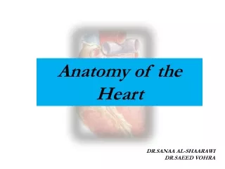
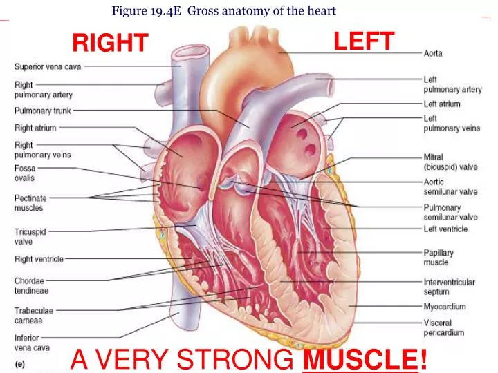
Figure 19.4E Gross anatomy of the heart. LEFT. RIGHT. A VERY STRONG MUSCLE !. Chambers of the heart blue=CO2, red =O2. Enters from body. Blood is deoxygenated, but has carbon dioxide. Comes back from lungs with oxygen. Valve. Valve. Valve. Valve. Goes out to the body with oxygen.

E N D
Figure 19.4E Gross anatomy of the heart LEFT RIGHT A VERY STRONG MUSCLE!
Chambers of the heartblue=CO2, red =O2 Enters from body. Blood is deoxygenated, but has carbon dioxide. Comes back from lungs with oxygen Valve Valve Valve Valve Goes out to the body with oxygen. Drops of oxygen and picks up carbon dioxide Goes to lungs. Drops off carbon dioxide and picks up oxygen.
Figure 19.5 Systemic & pulmonary circuits Blood from body enters Right Atrium Arteries carry blood AWAY from the heart Right Ventricle sends blood to lungs Veins carry blood to the heart (like ven in Spanish) Blood from lungs enters Left Atrium Left Ventricle sends blood to rest of body
N1WT/WT N1ΔFC/ΔFC A. E18.5 H&E B. E16.5 C. Mrs. Workman’s research!
N1WT/WT N1ΔFC/ΔFC Misformed valves!
Figure 19.6 Anatomical differences in the right & left ventricles Why is the left ventricle so much thicker than the right ventricle?
Valves • Prevent back flow • Between atria and ventricle-open when pressure increases due to a lot of blood in atria • Below ventricle-opens when ventricle contracts, pushing blood out through valve. • Slamming shut makes your heart beat. • What is a pulse?
Circulatory system • Works closely with system to bring oxygen from the lungs to the muscles and carbon dioxide from the muscles to the lungs to expel.
Blood Vessels • Arteries-move blood AWAY from the heart. Thicker walls because more pressure. • Vein-moves blood towards the heart. Thinner walls because less pressure. Valves to prevent back flow.
More on Blood Vessels • Capillaries-small blood vessels where gas exchange via diffusion takes place.