Download
1 / 59
590 likes | 796 Views
The Muscular System. Overview of Muscle Tissues. Muscles are responsible for all types of body movement Three basic muscle types are found in the body Skeletal muscle Cardiac muscle Smooth muscle. Functions of Skeletal Muscle. Produce movement Maintain posture Stabilize joints

E N D
Overview of Muscle Tissues • Muscles are responsible for all types of body movement • Three basic muscle types are found in the body • Skeletal muscle • Cardiac muscle • Smooth muscle
Functions of SkeletalMuscle • Produce movement • Maintain posture • Stabilize joints • Generate heat Hey Foxy Momma!
Characteristics of Muscles • Muscle cells are elongated (muscle cell = muscle fiber) • Contraction of muscles is due to the movement of microfilaments
Skeletal Muscle Characteristics • Most are attached by tendons to bones • Cells are multinucleate • Striated – have visible banding • Voluntary – subject to conscious control • Cells are surrounded and bundled by connective tissue
Connective Tissue Wrappings of Skeletal Muscle • Endomysium – around single muscle fiber • Perimysium – around a fascicle (bundle) of fibers • Epimysium – covers the entire skeletal muscle • Fascia – on the outside of the epimysium
Skeletal Muscle Attachments • Epimysium blends into a connective tissue attachment • Tendon – cord-like structure • Aponeuroses – • sheet-like structure • Sites of muscle attachment • Bones • Cartilages
Skeletal Muscle Subdivisions • Type I (Slow Twitch) (SO) (red) • low power production • high endurance capability • utilize fat breakdown for energy • generous blood supply • Type IIa (Mixed Characteristics) (FOG) • Type IIb (Fast Twitch) (FG) (white) • high power production • low endurance capability • utilize stored sugars/ATP for energy • low blood supply
Smooth Muscle Characteristics • Has no striations • Spindle-shaped cells • Single nucleus • Involuntary – no conscious control • Found mainly in the walls of hollow organs
Cardiac Muscle Characteristics • Has striations • Usually has a single nucleus • Joined to another muscle cell at an intercalated disc • Involuntary • Found only in the heart
Muscle Types: Cardiac Cross-Section
Microscopic Anatomy of Skeletal Muscle • Cells are multinucleate • Nuclei are just beneath the sarcolemma • Sarcolemma – specialized plasma membrane • Sarcoplasmic reticulum – specialized smooth endoplasmic reticulum
Microscopic Anatomy of Skeletal Muscle • Myofibril • Bundles of myofilaments (myosin specific microfilaments) • Myofibrils are aligned to give distinct bands • I band = light band • A band = dark band
Microscopic Anatomy of Skeletal Muscle • Sarcomere • Contractile unit of a muscle fiber • Organization of the sarcomere • Thick filaments = myosin filaments • Composed of the protein myosin • Has ATPase enzymes • Thin filaments = actin filaments • Composed of the protein actin
Microscopic Anatomy of Skeletal Muscle • Myosin filaments have heads (extensions, or cross bridges) • Myosin and actin overlap somewhat • At rest, there is a bare zone that lacks actin filaments • Sarcoplasmic reticulum (SR) – for storage of calcium
Skeletal Muscle Activity • Properties of Skeletal Muscle Activity • Irritability – ability to receive and respond to a stimulus • Contractility – ability to shorten when an adequate stimulus is received
Nerve Stimulus to Muscles • Skeletal muscles must be stimulated by a nerve to contract • Motor unit • One neuron • Muscle cells stimulated by that neuron • Neuromuscular junctions – association site of nerve and muscle • Synaptic cleft – gap between nerve and muscle • Nerve and muscle do not make contact • Area between nerve and muscle is filled with interstitial fluid
Transmission of Nerve Impulse to Muscle • Neurotransmitter – chemical released by nerve upon arrival of nerve impulse • The neurotransmitter for skeletal muscle is acetylcholine (Ach) • Neurotransmitter attaches to receptors on the sarcolemma • Sarcolemma becomes permeable to sodium (Na+) • Sodium rushing into the cell generates an action potential • Once started, muscle contraction cannot be stopped
The Sliding Filament Theory of Muscle Contraction • Activation by nerve causes myosin heads (crossbridges) to attach to binding sites on the thin filament • Myosin heads then bind to the next site of the thin filament • This continued action causes a sliding of the myosin along the actin • The result is that the muscle is shortened (contracted)
Brain initiates impulse to muscle Impulse reaches terminal end of axon Acetylcholine (Ach) is released across synaptic cleft Ach binds to sarcolemma Sarcolemma becomes permeable to Na+ and begins new action potential Action potential causes calcium (Ca 2+)to be released from sarcoplasmic reticulum Calcium binds to troponin on actin fiber causing myosin binding sites to be exposed ATP binds to myosin globular head Metabloism of ATP to ADP + free phosphate group causes globular head to change shape, grab onto myosin binding sites and “stroke” or pull actin fibers to the ‘M’ line of myosin, thus causing a muscle contraction. Calcium frees from troponin, ADP breaks off of myosin head and relaxion of muscle takes place Muscle Contraction Summary
Contraction of a Skeletal Muscle • Muscle fiber contraction is “all or none” • Within a skeletal muscle, not all fibers may be stimulated during the same interval • Different combinations of muscle fiber contractions may give differing responses • Graded responses – different degrees of skeletal muscle shortening
Types of Graded Responses • Twitch • Single, brief contraction • Not a normal muscle function • Tetanus (summing of contractions) • One contraction is immediately followed by another • The muscle does not completely return to a resting state • The effects are added
Types of Graded Responses • Unfused (incomplete) tetanus • Some relaxation occurs between contractions • The results are summed • Fused (complete) tetanus • No evidence of relaxation before the following contractions • The result is a sustained muscle contraction
Muscle Response to Strong Stimuli • Muscle force depends upon the number of fibers stimulated • More fibers contracting results in greater muscle tension • Muscles can continue to contract unless they run out of energy
Energy for Muscle Contraction • Initially, muscles used stored ATP for energy • Bonds of ATP are broken to release energy • Only 4-6 seconds worth of ATP is stored by muscles • After this initial time, other pathways must be utilized to produce ATP
Energy for Muscle Contraction • Direct phosphorylation • Muscle cells contain creatine phosphate (CP) • CP is a high-energy molecule • After ATP is depleted, ADP is left • CP transfers energy to ADP, to regenerate ATP • CP supplies are exhausted in about 20 seconds
Energy for Muscle Contraction • Aerobic Respiration • Series of metabolic pathways that occur in the mitochondria • Fatty acids & Glucose are broken down to carbon dioxide and water, releasing energy • This is a slower reaction that requires continuous oxygen
Energy for Muscle Contraction • Anaerobic glycolysis • Reaction that breaks down glucose without oxygen • Glucose is broken down to pyruvic acid to produce some ATP • Pyruvic acid is converted to lactic acid • This reaction is not as efficient, but is fast • Huge amounts of glucose are needed • Lactic acid produces muscle fatigue
Muscle Fatigue and Oxygen Debt • When a muscle is fatigued, it is unable to contract • The common reason for muscle fatigue is oxygen debt • Oxygen must be “repaid” to tissue to remove oxygen debt • Oxygen is required to get rid of accumulated lactic acid • Increasing acidity (from lactic acid) and lack of ATP causes the muscle to contract less
Types of Muscle Contractions • Isotonic contractions • Myofilaments are able to slide past each other during contractions • Concentric • Eccentric • Isometric contractions • Tension in the muscles increases • The muscle is unable to shorten • Static
Muscle Tone • Some fibers are contracted even in a relaxed muscle • Different fibers contract at different times to provide muscle tone • The process of stimulating various fibers is under involuntary control
Muscles and Body Movements • Movement is attained due to a muscle moving an attached bone • Muscles are attached to at least two points • Origin – attachment to a moveable bone • Insertion – attachment to an immovable bone
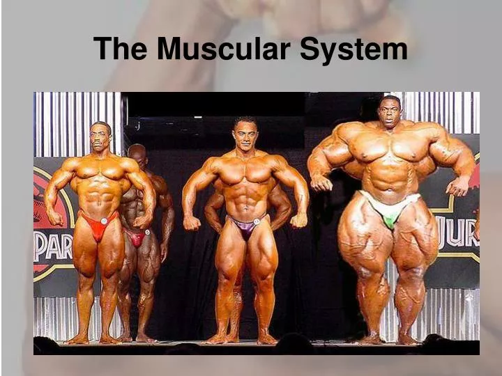
Effects of Exercise on Muscle • Results of increased muscle use • Increase in muscle size • Increase in muscle strength • Increase in muscle efficiency • Muscle becomes more fatigue resistant
Types of Ordinary Body Movements • Flexion • Extension • Rotation • Abduction • Circumduction
Special Movements • Dorsifelxion (toes to knees) • Plantar flexion (calve raise) • Inversion (foot in) • Eversion (foot out) • Supination (palm up) • Pronation (palm down) • Opposition (finger to finger)