Download
1 / 89
940 likes | 1.06k Views
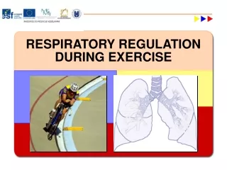
Respiration During Exercise. Objectives . Explain the principle physiological function of the pulmonary system. Outline the major anatomical components of the respiratory system. List major muscles involved in inspiration and expiration at rest and during exercise.

E N D
Objectives • Explain the principle physiological function of the pulmonary system. • Outline the major anatomical components of the respiratory system. • List major muscles involved in inspiration and expiration at rest and during exercise. • Discuss the importance of matching blood flow to alveolar ventilation in the lung. • Explain how gases are transported across the blood-gas interface in the lung.
Objectives • Discuss the major transportation modes of O2 and CO2 in the blood. • Discuss the effects of increasing temperature, decreasing pH, and increasing levels of 2–3 DPG on the oxygen-hemoglobin dissociation curve. • Describe the ventilatory response to constant-load, steady-state exercise. What happens to ventilation if exercise is prolonged and performed in a high-temperature/humid environment?
Objectives • Describe the ventilatory response to incremental exercise. What factors are thought to contribute to the alinear rise in ventilation at work rate above 50% to 70% of VO2 max? • Identify the location and function of chemoreceptors and mechanoreceptors that are thought to play a role in the regulation of breathing. • Discuss the neural-humoral theory of respiratory control during exercise.
Function of the Lung Structure of the Respiratory System Conducting Zone Respiratory Zone Mechanics of Breathing Inspiration Expiration Airway Resistance Mechanics of Breathing Diffusion of Gases Blood Flow to the Lung Outline • Control of Ventilation Ventilatory Regulation at Rest Respiratory Control Center Ventilatory Control During Submaximal Exercise Ventilatory Control During Heavy Exercise • Do the Lungs Adapt to Exercise Training? • Does the Pulmonary System Limit Maximal Exercise Performance? • Ventilation-Perfusion Relationships • O2 and CO2 Transport in Blood Hemoglobin and O2 Transport Oxyhemoglobin Dissociation Curve O2 Transport in Muscle O2 Transport in Blood • Ventilation and Acid-Base Balance • Ventilation and Blood-Gas Responses to Exercise Rest-to-Work Transition Prolonged Exercise in a Hot Environment Incremental Exercise
Introduction • Pulmonary respiration • Ventilation • Exchange of O2 and CO2 in the lungs • Cellular respiration • O2 utilization and CO2 production by the tissues • Purposes of the respiratory system during exercise • Gas exchange between the environment and the body • Regulation of acid-base balance during exercise
Function of the Lung Function of the Lung • Means of gas exchange between the external environment and the body • Replacing O2 • Removing CO2 • Regulation of acid-base balance • Ventilation • Mechanical process of moving air into and out of lungs • Diffusion • Random movement of molecules from an area of high concentration to an area of lower concentration
Function of the Lung In Summary • The primary function of the pulmonary system is to provide a means of gas exchange between the environment and the body. Further, the respiratory system plays an important role in the regulation of the acid-base balance during exercise.
Structure of the Respiratory System Structure of the Respiratory System • Organs • Nose and nasal cavities • Pharynx and larynx • Trachea and bronchial tree • Lungs • Alveoli • Site of gas exchange • Diaphragm • Major muscle of inspiration
Structure of the Respiratory System Structure of the Respiratory System • Lungs are enclosed by membranes called pleura • Visceral pleura • On outer surface of lung • Parietal pleura • Lines the thoracic wall • Intrapleural space • Intrapleural pressure is lower than atmospheric • Prevents collapse of alveoli
Structure of the Respiratory System Major Organs of the Respiratory System Figure 10.1
Structure of the Respiratory System Position of the Lungs, Diaphragm, and Pleura Figure 10.2
Conducting zone Conducts air to respiratory zone Humidifies, warms, and filters air Components: Trachea Bronchial tree Bronchioles Respiratory zone Exchange of gases between air and blood Components: Respiratory bronchioles Alveolar sacs Surfactant prevents alveolar collapse Structure of the Respiratory System Conducting and Respiratory Zones
Structure of the Respiratory System Conducting and Respiratory Zones Figure 10.3
Structure of the Respiratory System The Bronchial Tree Figure 10.4
Structure of the Respiratory System Type II Alveolar Cells Figure 10.5
Structure of the Respiratory System In Summary • Anatomically, the pulmonary system consists of a group of passages that filter air and transport it into the lungs where gas exchange occurs within tiny air sacs called alveoli.
Mechanics of Breathing Mechanics of Breathing • Movement of air occurs via bulk flow • Movement of molecules due to pressure difference • Inspiration • Diaphragm pushes downward, ribs lift outward • Volume of lungs increases • Intrapulmonary pressure lowered • Expiration • Diaphragm relaxes, ribs pulled downward • Volume of lungs decreases • Intrapulmonary pressure raised
Mechanics of Breathing The Mechanics of Inspiration and Expiration Figure 10.6
Mechanics of Breathing The Muscles of Respiration Figure 10.7
Mechanics of Breathing A Closer Look 10.1Respiratory Muscles and Exercise • Do respiratory muscles fatigue during exercise? • Historically believed that respiratory muscles do not fatigue during exercise • Current evidence suggests that respiratory muscles do fatigue during exercise • Prolonged (>120 minutes) • High-intensity (90–100% VO2 max) • Do respiratory muscle adapt to training? • Yes! • Increased oxidative capacity improves respiratory muscle endurance • Reduced work of breathing
P1 – P2 Airflow = Resistance Mechanics of Breathing Airway Resistance • Airflow depends on: • Pressure difference between two ends of airway • Resistance of airways • Airway resistance depends on diameter • Chronic obstructive lung disease • Asthma and exercise-induced asthma
Mechanics of Breathing Clinical Applications 10.1Exercise-Induced Asthma • Asthma results in bronchospasm • Narrowing of airways • Increased work of breathing • Shortness of breath (dyspnea) • Many potential causes • Exercise-induced asthma • During or immediately following exercise • May impair exercise performance
Mechanics of Breathing Clinical Applications 10.2Exercise and Chronic Obstructive Lung Disease • Chronic obstructive lung disease (COPD) • Increased airway resistance • Due to constant airway narrowing • Decreased expiratory airflow • Includes two lung diseases: • Chronic bronchitis • Excessive mucus blocks airways • Emphysema • Airway collapse and increased resistance • Increased work of breathing • Leads to shortness of breath • May interfere with exercise and activities of daily living
Mechanics of Breathing In Summary • The major muscle of inspiration is the diaphragm. Air enters the pulmonary system due to intrapulmonary pressure being reduced below atmospheric pressure (bulk flow). At rest, expiration is passive. However, during exercise, expiration becomes active, using muscles located in the abdominal wall (e.g., rectus abdominis and internal oblique). • The primary factor that contributes to airflow resistance in the pulmonary system is the diameter of the airway.
Pulmonary Ventilation Pulmonary Ventilation • The amount of air moved in or out of the lungs per minute (V) • Tidal volume (VT) • Amount of air moved per breath • Breathing frequency (f) • Number of breaths per minute • Alveolar ventilation (VA) • Volume of air that reaches the respiratory zone • Dead-space ventilation (VD) • Volume of air remaining in conducting airways V = VT x f V = VA + VD
Pulmonary Ventilation In Summary • Pulmonary ventilation refers to the amount of gas moved into and out of the lungs. • The amount of gas moved per minute is the product of tidal volume times breathing frequency.
Pulmonary Volumes and Capacities Pulmonary Volumes and Capacities • Vital capacity (VC) • Maximum amount of gas that can be expired after a maximum inspiration • Residual volume (RV) • Volume of gas remaining in lungs after maximum expiration • Total lung capacity (TLC) • Amount of gas in the lungs after a maximum inspiration.
Pulmonary Volumes and Capacities Definitions of Pulmonary Volumes and Capacities
Pulmonary Volumes and Capacities Lung Volumes and Capacities Figure 10.9
Pulmonary Volumes and Capacities Spirometry • Measurement of pulmonary volumes and rate of expired airflow • Useful for diagnosing lung diseases • Chronic obstructive lung disease (COPD) • Spirometric tests • Vital capacity (VC) • Maximal volume of air that can be expired after maximal inspiration • Forced expiratory volume (FEV1) • Volume of air expired in 1 second during maximal expiration • FEV1/VC ratio • ≥80% is normal
Pulmonary Volumes and Capacities A Computerized Spirometer Figure 10.8
Pulmonary Volumes and Capacities Forced Expiratory Airflow Used to Diagnose Airway Obstruction Figure 10.10
Pulmonary Volumes and Capacities In Summary • Pulmonary volumes can be measured using spirometry. • Vital capacity is the maximum amount of gas that can be expired after a maximal inspiration. • Residual volume is the amount of gas left in the lungs after a maximal expiration.
Diffusion of Gases Partial Pressure of Gases • Dalton’s law • The total pressure of a gas mixture is equal to the sum of the pressure that each gas would exert independently • Calculation of partial pressure Pair = PO2 + PCO2 + PN2
A V gas = x D x (P1 – P2) T Diffusion of Gases Diffusion of Gases • Fick’s law of diffusion • The rate of gas transfer (V gas) is proportional to the tissue area, the diffusion coefficient of the gas, and the difference in the partial pressure of the gas on the two sides of the tissue, and inversely proportional to the thickness. V gas = rate of diffusion A = tissue area T = tissue thickness D = diffusion coefficient of gas P1 – P2 = difference in partial pressure
Diffusion of Gases Partial Pressures of O2 and CO2 and Gas Exchange Figure 10.11
Diffusion of Gases In Summary • Gas moves across the blood-gas interface in the lung due to simple diffusion. • The rate of diffusion is described by Fick’s law, which states: the volume of gas that moves across a tissue is proportional to the area for diffusion and the difference in partial pressure across the membrane, and is inversely proportional to membrane thickness.
Blood Flow to the Lung Blood Flow to the Lung • Pulmonary circuit • Same rate of flow as systemic circuit • Lower pressure • When standing, most of the blood flow is to the base of the lung • Due to gravitational force • During exercise, more blood flow to apex
Blood Flow to the Lung The Pulmonary and Systemic Circulation Figure 10.12
Blood Flow to the Lung Regional Blood Flow within the Lung Figure 10.13
Blood Flow to the Lung In Summary • The pulmonary circulation is a low-pressure system with a rate of blood flow equal to that in the systemic circuit. • In a standing position, most of the blood flow to the lung is distributed to the base of the lung due to gravitational force.
Ventilation-Perfusion Relationships Ventilation-Perfusion Relationships • Ventilation/perfusion ratio (V/Q) • Indicates matching of blood flow to ventilation • Ideal: ~1.0 • Apex of lung • Underperfused (ratio <1.0) • Base of lung • Overperfused (ratio >1.0) • During exercise • Light exercise improves V/Q ratio • Heavy exercise results in V/Q inequality
Ventilation-Perfusion Relationships Ventilation/Perfusion Ratios Figure 10.14
Ventilation-Perfusion Relationships In Summary • Efficient gas exchange between the blood and the lung requires proper matching of blood flow to ventilation (called ventilation-perfusion relationships). • The ideal ratio of ventilation to perfusion is 1.0 or slightly greater, since this ratio implies a perfect matching on blood flow to ventilation.
O2 and CO2 Transport in Blood O2 Transport in the Blood • 99% of O2 is transported bound to hemoglobin (Hb) • Oxyhemoglobin: Hb bound to O2 • Deoxyhemoglobin: Hb not bound to O2 • Amount of O2 that can be transported per unit volume of blood is dependent on the Hb concentration • Each gram of Hb can transport 1.34 ml O2 • Oxygen content of blood (100% Hb saturation) • Males: • Females: 150 g Hb/L blood x 1.34 ml O2/g Hb = 200 ml O2/L blood 130 g Hb/L blood x 1.34 ml O2/g Hb = 174 ml O2/L blood
O2 and CO2 Transport in Blood Oxyhemoglobin Dissociation Curve • Deoxyhemoglobin + O2 Oxyhemoglobin • Direction of reaction depends on: • PO2 of the blood • Affinity between Hb and O2 • At the lung • High PO2 = formation of oxyhemoglobin • At the tissues • Low PO2 = release of O2 to tissues
O2 and CO2 Transport in Blood Oxygen-Hemoglobin Dissociation Curve Figure 10.15
O2 and CO2 Transport in Blood Effect of pH, Temperature, and 2–3 DPG on the O2-Hb Dissociation Curve • pH • Decreased pH lowers Hb-O2 affinity • Results in a “rightward” shift of the curve • Favors “offloading” of O2 to the tissues • Temperature • Increased blood temperature lowers Hb-O2 affinity • Results in a “rightward” shift of the curve • 2–3 DPG • Byproduct of RBC glycolysis • May result in a “rightward” shift of the curve • During altitude exposure • Not a major cause of rightward shift during exercise
O2 and CO2 Transport in Blood Effect of pH on the Oxygen-Hemoglobin Dissociation Curve Figure 10.15