Download

1 / 18
180 likes | 379 Views
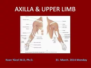
Anatomy of the hand. IN 14 QUESTIONS. Kaan Yücel M.D., Ph.D. 9.January.2013 Wednesday. 1. ...bones of the hand ?. 2. What are flexor retinaculum & carpal tunnel?. The carpal tunnel formed anteriorly at wrist by a deep arch formed by carpal bones & f lexor retinaculum

E N D
Anatomy of the hand • IN 14 QUESTIONS • Kaan Yücel M.D., Ph.D • 9.January.2013 Wednesday
2. What are flexor retinaculum & carpal tunnel? The carpal tunnelformed anteriorly at wrist by a deep arch formed by carpal bones & flexor retinaculum (transverse carpal ligament) Flexordigitorumsuperficialis Flexordigitorumprofundus Flexorpollicis longus Median nerve Pass through the carpal tunnel
3. What is extensor retinaculum (dorsal carpal ligament)? The extensor tendons pass into the hand in six compartments defined by an extensor retinaculum: • extensor digitorum & extensor indicis posterior surface of the wrist extensor carpi ulnaris & extensor digiti minimi medial side of the wrist • abductor pollicis longus & extensor pollicis brevis • extensor carpi radialis longus & extensor carpi radialis brevis • extensor pollicis longus • through three compartments on the lateral surface of the wrist.
4. What is palmar aponeurosis? A triangular condensation of deep fascia that covers the palm and is anchored to the skin in distal regions. Continuous with the palmaris longus tendon, when present; otherwise, anchored to the flexor retinaculum.
5. …Fibrousdigitalsheaths? • Tendonsof flexor digitorumsuperficialis and profunduscross the palm • enter fibrous sheaths on the palmar aspect of the digits. • formed by fibrous arches and cruciate (cross-shaped) ligaments • hold the tendons to the bony plane • prevent the tendons from bowing when the digits are flexed. are surrounded by a synovial
6. What are extensor hoods for? Tendons of the extensor digitorumextensor pollicis longus musclesexpand over the proximal phalanges to form "extensor hoods" or "dorsal digital expansions". Tendons of the extensor digiti minimi, extensor indicis, extensor pollicis brevisjoin these hoods.
7. Which are the intrinsic muscles of the hand? Palmaris brevis Thenar muscles Abductor pollicis brevis Abductor digiti minimi Opponens pollicis Hypothenar muscles Flexor digiti minimi Adductor pollicis Flexor pollicis brevis Lumbrical Interossei
8. …functionsthe intrinsic muscles of the hand? Palmarinterossei adduct the thumb, index, ring, and little fingers with respect to a long axis through the middle finger Dorsalinterossei major abductors of the index, middle, and ring fingers, at the metacarpophalangeal joints
8. …functionsthe intrinsic muscles of the hand? Lumbricals flexing metacarpophalangealjoints extending interphalangeal joints medial two deep branch of the ulnar nerve lateral two median nerve
8. …functionsthe intrinsic muscles of the hand? Palmarisbrevis deepens cup of the palm by pulling on skin over the hypothenar eminence forming a distinct ridge. This may improve grip.
9. How are the intrinsic muscles innervated? All of the intrinsic muscles of the hand by deep branch of the ulnar nerve Except three thenar & two lateral lumbrical muscles by median nerve
10. ....arteries of the hand? Superficial palmar arch Deep palmar arch
11....veins of the hand? Cephalic veinoriginates from lateral side of dorsal venous network. Basilic vein originates from medial side of dorsal venous network.
12. ...sensory innervation of the hand? Ulnar nerve medial side of the palm,medial half of the dorsum of the hand, the 5th finger, and the medial half of the 4th finger,anterior surfaces of the medial one and a half digits, Median nerve thumb,index,middle fingers,lateral side of the ring [distal parts on the dorsum of the hand] Radial nerve dorsolateral side
13. ...carpal tunnel? base of the carpal arch formed medially by pisiform & hook of hamate laterally by tubercles of scaphoid & trapezium SLTP TTCH
14. ....anatomical snuffbox? • lateral border • tendons of • abductor pollicislongusAPLEPB • extensor pollicisbrevis • medial border • tendon of extensor pollicislongus
14. ....anatomical snuffbox? • floor • scaphoid & trapezium • distal ends of the tendons of extensor carpi radialislongusextensor carpi radialisbrevis