Download
1 / 15
150 likes | 324 Views
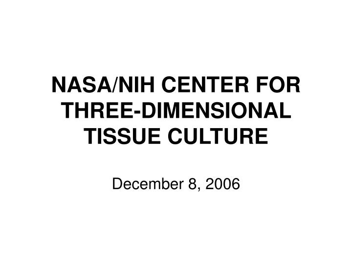
NASA/NIH CENTER FOR THREE-DIMENSIONAL TISSUE CULTURE. December 8, 2006. NASA/NIH Center for Three-Dimensional Tissue Culture. Brings the technology of the NASA-rotating wall vessel bioreactor to the NIH community Mutual leverage of research across Agencies Peer Review by NICHD

E N D
NASA/NIH CENTER FOR THREE-DIMENSIONAL TISSUE CULTURE December 8, 2006
NASA/NIH Center for Three-Dimensional Tissue Culture • Brings the technology of the NASA-rotating wall vessel bioreactor to the NIH community • Mutual leverage of research across Agencies • Peer Review by NICHD • Microgravity Incubator for ISS science • Facilities for scientists to: • launch pilot projects on the use of the bioreactor for their research interest • continue those projects by submitting small grant proposals • Intellectual support for phase III projects
PROJECTS AT THE NASA/NIH CENTER • HIV pathogenesis in human lymphoid tissue. • Repopulation of thymus tissue. • Study of hormonal regulation in pineal gland. • Study of nerve interactions in brain tissue slices. • Model of atherosclerosis. • Osmotic regulation in renal tubules. • Follicular development in mammmalian ovary. • Differentiation of mesangial cells. • Role of laminin in differentiation of salivary gland. • Model of T cell differentiation and traffic. • Study of progression of metaplasia to dysplasia and carcinoma. • Long term maintenance of human prostate tissues. • Models of adenocarcinoma invasion/metastasis. • Hepatocyte infection by avain malaria. • Production of Herpes Zoster Virus in skin explants. • Improving vector transfer in gene therapy. • Protein crystal growth. • Replication of Cyclospora in intestinal cells. • Study of neurosecretory pathways in rat hypothalamus. • Study of mucolipidosis type 4 in epithelial cells. • Differentiation of dendritic cells. • Role of gap junctions in lymphoid tissue. • Characterization of tonsil T cell response to RSV • Hematopoeitic stem cell culture for stem cell therapy in space • Model for pathogenesis of Kaposi’s sarcoma. • Development of advanced dual-photon microscope. • Improving activity of mononuclear phagocytes. • Uterine fibroid genesis and progression. • Maintenance of preneoplastic lesions of the lung. • Developing a “universal” pathogen culture system. • Effect of microgravity on the immune function of human lymphoid tissues. • Study of normal and abnormal endometrium. • Culture of Hermanski-Pudlack Syndrome fibroblasts. • Modeling of Lyme disease. • Culture of esophageal carcinoma and gastrinoma. • Culture of fetal islet cells for diabetes research. • Whole mouse embryo culture. • Culture of synovial tissues from arthritic patients. • Cryptosporidium infection of intestinal cells. • Study of HIV mucosal transmission. • Growth of Norwalk-like viruses. • Lens cell culture for study of cataracts. • Culture of hematopoietic progenitor cells for gene therapy of hemoglobinopathies. • GM-CSF activity in tumor cells. • Neurogenesis in a cell-hydrogel system • Urogenital and intestinal epitelial models of pathogenic E. Coli • Extracellular signals ondifferentiation of embryonic stem cells • Mechanism of malaria parasite culture and release • Culture of normal and metastatic breast stem cells
NASA/NIH Center in NICHD Leonid Margolis The development of the first system of human lymph node and tonsillar tissues ex vivo, that preserves tissues structure and immune function. This system is currently used by various groups to study HIV pathogenesis. Based on this work, other groups in collaboration with the NASA/NIH Center developed an ex vivo system of gut-associated lymphoid tissues – the main target for HIV-1. Also, ex vivo system of cervical tissue to study HIV transmission to women has been developed. These ex vivo tissue systems are now used by laboratories in the USA and Europe to study HIV pathogenesis and to test anti-HIV compounds. • As the result of this work two large NIH-funded programs have been designed and are currently funded by NIH: • A multi-laboratory microbicide quality assurance program (MQAP) aimed to standardize the ex vivo tissue systems. • An international Program (U19) for pre- clinical evaluation of microbicides efficacyin tissue ex vivo.
Cultured lymphoid tissue blocks retain their cytoarchitecture T cells B cells Macrophage FDC Network
500 18000 RWV RWV Static Static 15000 400 12000 300 Ig M (ng/ml) Anti-TT IgG (mU/ml) 9000 200 6000 100 3000 0 0 3 6 9 12 15 18 3 6 9 12 15 18 days in culture days in culture Human lymphoid tissues respond to recall antigen challenge or polyclonal stimulation but not in RWV
35 Activated 30 25 20 Antibody Production Index 15 10 5 0 Static ISS Effect of microgravity on antibody production
Activated condition Pre-activated condition 7000 1600 P1 P1 1400 6000 P3 P3 P4 Ig (ng/ml) 1200 5000 P5 P5 Ig (ng/ml) 1000 4000 800 3000 600 2000 400 1000 200 0 0 Ground ACT Flight ACT Ground PreACT Flight PreACT ISS x4: antibody production - individual patients
ACT/NA Ground Flight p IFN-g33.43+13.67 1.86+0.74 0.15 IL-2 2.07+1.04 1.01+0.28 0.32 TNF-a 499.35+311.25 1.16+0.27 0.25 IL-6 5.93+1.18 0.58+0.20 0.04 IL-1a 2.30+0.59 0.90+0.15 0.09 IL-8 3.48+0.65 1.18+0.43 0.01 IL-16 1.55+0.03 0.99+0.18 0.11 MIP-1b 27.20+1.31 1.06+0.20 0.00 RANTES 75.54+6.36 1.16+0.11 0.01 SDF-1 10.08+2.40 1.04+0.19 0.06 preACT/NA Ground Flight p IFN-g11.19+5.54 2.31+0.61 0.20 IL-2 1.62+0.34 1.12+0.22 0.43 TNF-a 162.15+67.71 46.37+33.36 0.08 IL-6 1.26+0.46 0.49+0.17 0.31 IL-1a 9.87+5.96 4.26+2.32 0.22 IL-8 0.92+0.33 0.56+0.23 0.50 IL-16 1.50+0.24 0.99+0.10 0.06 RANTES 79.92+22.81 31.97+11.21 0.01 SDF-1 29.38+19.23 23.50+12.85 0.59 Four cytokines/chemokines, IL-6, IL-8, MIP1b, and RANTES were significantly lower in flight activated conditions compared to ground controls. None were significantly different in pre-activated conditions. ISS x4: Cytokine/chemokine secretion
CBOSS-FDI: Bubble removal and good mixing on ISS with cell culture bags and cellular analogs Before Removal and Mixing %CV Map Final Result – Well Mixed with few Bubbles Mean %CV = 5% % Bubbles = 1% After Removal and Mixing Digital Removal
Repopulation Of Tonsillar Tissue in RWV With Labeled Lymphocytes
NICHD support for NASA intramural scientists • Version 2.1.2.0 • Abstract • Grant Number: 1R03HD044557-01PI Name: REINSCH, SIGRID S.PI Email: sreinsch@mail.arc.nasa.gov PI Title: GROUP LEADERProject Title: Homologous recombination in Xenopus and Zebrafish • Abstract: DESCRIPTION (provided by applicant): The long-term objective of these studies is to generate novel methodologies for genetic manipulation of vertebrate embryos for the ultimate purpose of understanding gene function during development. The investigators propose experiments to test the hypothesis that bacteriophage protein pairs can stimulate homologous recombination (HR) in vertebrate Xenopus and Zebrafish embryos. The bacteriophage protein pairs include Red__ and Red__ from phage lambda, and RecE and RecT from the lambdoid prophage Rac. The protein pairs have been employed for both efficient gene conversion and gap repair using short homology regions. Recombination involves co-operation between a 5'-3' exonuclease (RecE or Red-alpha with a single strand binding protein (RecT or Red-Beta). Our collaborator, A.F. Stewart, pioneered the use of these proteins to initiate homologous recombination in E. coli. Recently Dr. Stewart's lab has demonstrated that these proteins can promote homologous recombination in mouse ES cells using single stranded oligonucleotides to target specific sequences. This has been termed ssOR for single-strand oligo repair. This exploratory grant will investigate several approaches in early Xenopus or Zebrafish embryos to target specific chromosomal sequences using the phage protein pairs to promote ssOR. The investigators propose to (1) develop assays for ssOR in cytoplasmic extracts of Xenopus eggs to explore the specific requirements for HR in the Xenopus embryonic cytoplasm. The assay will examine requirements for repair of antibiotic resistance genes on extrachromosomal plasmids and will use an E.coli colony readout. (2) The investigators will target a chromosomal Green Fluorescent Protein (GFP) transgene in Xenopus tropicalis embryos to generate visually screenable loss of function phenotypes. Purified phage proteins and single strand oligos will be used target a GFP transgene to alter the expression of GFP expression through coding changes, frameshift mutation, or stop codon insertion. (3) The investigators will explore conditions to target several zebrafish chromosomal loci using ssOR to generate well-characterized point mutations that give scorable phenotypes. • Thesaurus Terms:Xenopus, developmental genetics, gene targeting, zebrafish virus protein bacterial virus, biotechnology, embryo /fetus, embryonic stem cell, genetically modified animal, green fluorescent protein, oligonucleotide, point mutation Institution: NASA--AMES RESEARCH CENTER241-1MOFFETT FIELD, CA 940351000Fiscal Year: 2004Department: Project Start: 01-JAN-2004Project End: 31-DEC-2005ICD: NATIONAL INSTITUTE OF CHILD HEALTH AND HUMAN DEVELOPMENTIRG: CHHD