Download
1 / 19
810 likes | 5k Views
The Lymphatic System. Dr. Mustafa Saad (2018). Lymphatic System S tructure and Function. Consists of: Lymph Lymphatic vessels Structures and organs containing lymphatic tissue Red bone marrow. Functions of the lymphatic system Drains excess interstitial fluid.

E N D
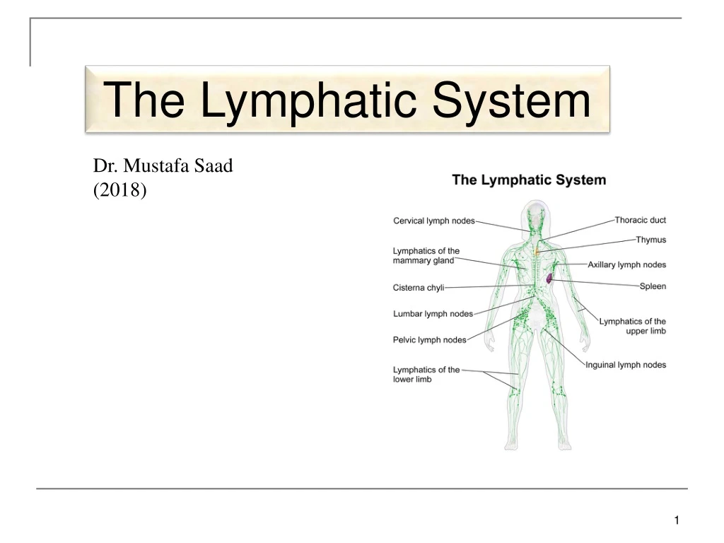
The Lymphatic System Dr. Mustafa Saad (2018)
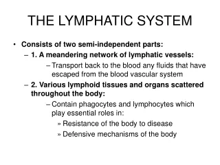
Lymphatic System Structure and Function • Consists of: • Lymph • Lymphatic vessels • Structures and organs containing lymphatic tissue • Red bone marrow • Functions of the lymphatic system • Drains excess interstitial fluid. • Transports dietary lipid from gastrointestinal tract to blood. The lipid-containing lymph is called Chyle. • Carry out immune responses. Fig.1: Components of the lymphatic system.
Lymph • Most components of blood plasma filter freely through the capillary walls to form interstitial fluid. • More fluid filters out of blood capillaries than returns to them by reabsorption. • The excess filtered fluid drains into lymphatic vessels and becomes lymph, Fig.2: Capillaries and lymphatics.
Lymphatic Vessels • Vessels begin as lymphatic capillaries. These are closed at one end. • Lymphatic capillaries unite to form large lymphatic vessels. These resemble veins in structure but thinner walls and more valves. • A series of swellings are present along the lymphatic vessels. These are the lymph nodes. Fig.3: The distribution of lymphatic vessels in the body showing the lymph nodes. Also shown is the closed end of a lymphatic capillary.
Ultimately, the lymph drains into 2 main channels: • Thoracic duct: a long duct that drains lymph from the entire left half of the body and the right half below the ribs including the right lower limb. • Right lymphatic duct: a short duct that drains lymph from the right side of the body above the ribs including the right upper limb. Fig.4: Areas of lymph drainage to the two main lymphatic ducts.
Each of these ducts open into the junction of the subclavian and internal jugular veins as they form the brachiocephalic vein. • In this way, the lymph will return to the blood. Fig.5: Opening of the lymphatic ducts into the venous circulation.
Lymphatic Organs and Tissues • 2 groups based on function • Primary lymphatic organs: • Sites where B and T lymphocytes are formed and mature. • Red bone marrow and thymus. • Secondary lymphatic organs • Sites where most immune response occurs, meaning that they are the sites where the mature lymphocytes perform their function. • Lymph nodes, spleen, lymphatic nodules.
The Thymus • The thymus is an asymmetric bilobed organ where mature T-cells are formed. • It’s located in the superior mediastinum just behind the manubrium. It may descend into the anterior mediastinum to lie between the sternum and the pericardial sac. Sometimes it may ascend into the neck reaching as high as the thyroid gland. • The fibrous capsule that surrounds the gland sends connective tissue trabeculae into the gland dividing it into lobules. Each lobule is formed of a dark outer region with immature T-cells (the cortex) and an inner lighter region with mature T-cells (the medulla).
Fig.6: To the left, histology of the thymus. Below, position of the thymus. • The thymic tissue is most numerous in younger age. As the person grows, this tissue is gradually replaced by fatty tissue.
Red Bone Marrow • Red bone marrow is the site of formation of the blood elements: red blood cells, white blood cells and platelets. • Inside the red bone marrow B lymphocyte form and mature; T lymphocytes are formed in the red bone marrow but they’re immature. T-cells become mature in the thymus. • Red bone marrow in adults is present in the flat bones and the epiphyses of some long bones.
Lymph Nodes • Lymph nodes are capsulated bean shaped structures that are found along the course of the lymphatic vessels. They’re usually present in groups and they’re scattered all over the body. • The capsule sends trabeculae into the node dividing it into compartments. Beneath the capsule there’s a space called the Subcapsular Sinus. • The node has an outer cortex with lymphatic nodules formed of B-cells and plasma cells. Deep to it is the inner cortex formed of T-cellswith no nodules. Deep to the cortex is the medulla which contains B-cells, plasma cells and macrophages embedded in reticular fibers. • Between the lymphatic tissue in the cortex and medulla are spaces called cortical and medullary sinuses.
Important Note: • In any organ in the body, the tissues forming the organ can be divided into two types: • The Parenchyma: any tissue in the organ responsible for performing the function of the organ. Example: in the lymph nodes, the lymphoid tissue is the parenchyma. • The Stroma: tissues in the organ that are not related to the function of the organ but play a supporting role. Example: in the lymph nodes, the capsule, the trabeculae, the reticular fibers and fibroblasts are the stroma.
From the convex side of the lymph node several incoming lymphatic vessels enter the node. These are called afferent lymphatic vessels. • The concave side of the node is called the hilum. From it, one or two outgoing lymphatic vessels leave the node. These are called efferent lymphatic vessels. Also through the hilum arteries and nerves enter and veins exit the node. Medullary Sinuses • Efferents Cortical Sinuses Afferents Subcapsular Sinus The flow of lymph in a lymph node
Functions of lymph nodes: Filter of lymph • Antigens in the lymph are trapped and the lymphocytes in the nodes react to it and initiate the immune response. • Macrophages in the node may directly destroy the antigen. • It’s important to know what lymphatic vessels drain a certain organ and what are the lymph nodes in its course, because this represent a pathway by which infections and cancer cells can spread.
The Spleen • Largest single mass of lymphatic tissue in the body. • It’s an oval, soft organ located in the left hypochondriac region. • Its superior surface is smooth and related to the diaphragm and ribs 9,10 and 11. Its visceral surface is irregular and related to the stomach, pancreas, kidney and colon. • Stroma – capsule, trabeculae, reticular fibers, and fibroblasts. • Parenchyma: • White pulp – lymphatic tissue (lymphocytes and macrophages) surrounds branches of splenic artery (the artery that supplies the spleen and enters through the hilum).
Red pulp – blood-filled venous sinuses surrounded by splenic cords which contain red blood cells, macrophages, lymphocytes and plasma cells. • Macrophages remove ruptured, worn out or defective blood cells. • Storage of up to 1/3 of body’s platelet supply. • Production of blood cells during fetal life. • Function: Filter of Blood
Lymphatic Nodules • Collection of lymphatic tissue not surrounded by a capsule. • Scattered throughout lining of gastrointestinal, urinary, reproductive and respiratory tracts. • Most of them are small and solitary. • Some are large – tonsils, Peyer’s patches, appendix.