Download
1 / 29
290 likes | 610 Views
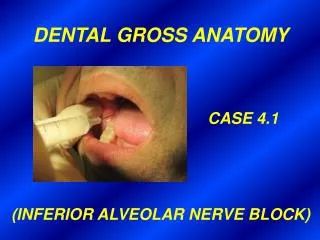
DENTAL GROSS ANATOMY CASE 2.1. History A 36yo woman slept near an open window on a cold drafty night. Upon awakening she noticed that her face was distorted. She was not able to close her right eye and she had difficulty speaking, eating (but not

E N D
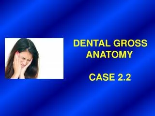
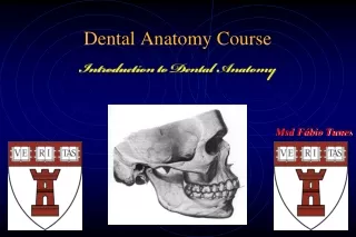
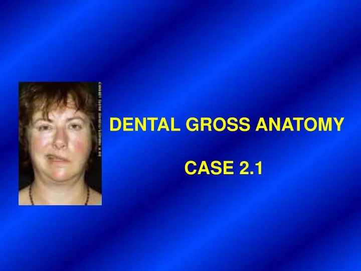
DENTAL GROSS ANATOMY CASE 2.1
History • A 36yo woman slept near an open window on • a cold drafty night. • Upon awakening she noticed that her face • was distorted. • She was not able to close her right eye and • she had difficulty speaking, eating (but not • swallowing) and drinking. Additionally, her • sense of taste was impaired. • She consulted her physician.
Examination • Examination reveals that the patient’s face is • immobile and without expression on the right • side. • Right forehead is without wrinkles and right • eyebrow droops. • Right lower eyelid sags and tears flow down • her face. • Right corner of her mouth sags, and she cannot • purse her lips. • Nose and mouth are deviated toward the • unaffected side. • When attempting to laugh, the facial distortion • becomes more noticeable.
Diagnosis, Therapy and Further Course • A diagnosis was made. • Therapy included electrical stimulation, • massage and active exercises of the facial • muscles. • After five weeks the patient was almost • completely recovered and only traces of the • paralysis could be seen.
1a. What is the diagnosis? b. What major structure was affected?
PEOPLE AFFLICTED WITH BELL’S PALSY (DAMAGE TO FACIAL NERVE)
What is (are) the underlying • cause(s) of this condition?
VII. FACIAL N. VII entering internal auditory meatus VII in facial canal of temporal bone
Posterior auricular a. ( supplies VII) VII
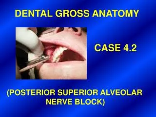
3a. What is the name of the foramen by which this major structure leaves the base of the skull? b. Name the five terminal branches of this structure.
Stylomastoid foramen (exit of VII)
Temporal branches Zygomatic branches Posterior auricular n. (to occipitalis m.) Main trunk of VII TO ZANZIBAR BY MOTOR CAR Buccal branches Marginal mandibular branch Cervical branch
4a. Why were wrinkles absent from the patient’s forehead and why did the right eyebrow droop? b. Why was the patient unable to purse her lips and why did liquids run out of the corner of her mouth when she tried to drink?
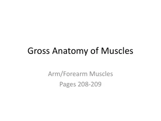
Frontalis m. Orbicularis oris m.
4c. Paralysis of which muscle resulted in food collecting in the vestibule of the patient’s mouth? What is the anatomical origin of this muscle? What is its embryological origin? Is it a true muscle of mastication?
Pterygoid hamulus Buccinator m. Pterygomandibular raphe Superior pharyngeal constrictor m. Mandible Buccinator also originates from alveolar processes of maxilla and mandible
PHARYNGEAL ARCH MUSCLES V3 VII IX X X Muscles of mastication: Arch 1 Muscles of facial expression: Arch 2 (INCLUDING BUCCINATOR)
4d. Why couldn’t the patient close her right eye? What serious complication might be the result?
Orbicularis oculi m. (orbital part) Orbicularis oculi m. (palpebral part)
In this patient there was sagging • of the lower eyelid and spilling of • tears down the side of her face. • What is the pathway by which • tears normally drain from the • conjunctival sac?
Superior lacrimal papilla and punctum Lacrimal gland (orbital part) Lacrimal canaliculi Lacrimal gland (palpebral part) Lacrimal sac Ducts of lacrimal gland (open into superolateral part of conjunctival sac) Nasolacrimal duct Opening of nasolacrimal duct (into inferior meatus of nasal cavity) Inferior lacrimal papilla and punctum
6a. Why did this patient experience impairment of taste? b. Where, precisely, was the loss of taste sensation?
COURSE OF VII IN FACIAL CANAL Geniculate ganglion (sensory) Int. auditory meatus Greater petrosal n. (autonomic to lacrimal gland) VII in facial canal of temporal bone Nerve to stapedius m. (paralysis > hyperacusis) Pterygopalatine ganglion (autonomic) Stylomastoid for. Chorda tympani n. (autonomic to submandibular & sublingual glands, taste from ant. 2/3 of tongue) Submandibular ganglion (autonomic) Branches to muscles of facial expression
Additional note: According to Moore & Agur (Essential Clinical Anatomy), VII is the most frequently paralyzed cranial nerve.