Download
1 / 55
550 likes | 658 Views
Chapter 8 The Special Senses. Biology 112 Tri-County Technical College Pendleton, SC. You’re special…I’m special. Usually told we have five senses: touch, taste, smell, sight, and hearing

E N D
Chapter 8 The Special Senses Biology 112 Tri-County Technical College Pendleton, SC
You’re special…I’m special • Usually told we have five senses: touch, taste, smell, sight, and hearing • TOUCH actually mixture of general senses: temperature, pressure, and pain receptors of skin and proprioceptors of muscles and joints • Smell, taste, sight, and hearing are called special senses
Special Senses, cont. • Receptors for 5th sense, equilibrium, are housed in ear along with organ of hearing • Special sense receptors are either large, complex sensory organs (such as eyes and ears) or localized clusters of receptors (such as taste buds and olfactory epithelium)
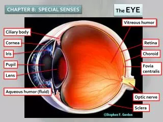
The Human Eye • Eyelids meet at medial and lateral canthus (protection) • Modified sebaceous glands at eyelid edges called meibomian glands produce oily secretion that lubricates eyes • Ciliary glands (modified sweat glands) lie between eyelashes and help lubricate eyeball • Sty is inflammation of one of these ciliary glands
Human Eye, cont. • Conjunctiva is delicate membrane that lines eyeball and covers parts of outer surface of eyeball • Ends at edge of cornea by fusing with corneal epithelium • Secretes mucus to lubricate and keep eyeball moist • Conjunctivitis (inflammation of conjunctive) results in reddened, irritated eyes • Pinkeye (conjunctiva infection caused by bacteria/virus) is highly contagious
Human Eye III • Lacrimal apparatus consists of number of glands and ducts that drain lacrimal secretions into nasal cavity • Lacrimal glands located above lateral end of each eye release dilute salt solution (tears) onto anterior surface of eyeball through lacrimal gland ducts
Human Eye IV • Tears flush across eyeball into lacrimal canals medially, then into lacrimal sac, and finally into nasolacrimal duct which empties into nasal cavity • Tears contain antibodies and lysozyme • Cleanses and protects as moistens and lubricates • Sclera (thick, white connective tissue) is outermost protective tunic (also called fibrous tunic • Seen anteriorly as “white of the eye”
Human Eye V • Central anterior portion (sclera) modified to crystal clear • This transparent window is called the cornea allows light to enter the eye • Iris is pigmented part of eye and contains the pupil • Six extrinsic (external) eye muscles attached to outer surface of each eye • Produce gross eye movements and make it possible for eye to follow a moving object
Eye Tunics • Eye (eyeball) is hollow sphere whose wall is composed of three tunics (coats) • Its interior is filled with fluids called humors that help maintain its shape • Outermost tunic (sclera or fibrous tunic) is thick, white connective tissue seen anteriorly as the “white of the eye” • Central anterior portion modified to crystal clear structure called the cornea through which light enters eye
Eye Tunics, cont. • Cornea well supplied with nerve endings, has ability to repair itself • **Cornea has NO blood vessels and is ONLY tissue that can be transplanted from one person to another without fear of rejection • Choroid is middle coat of eyeball and is blood-rich nutritive tunic containing dark pigment
Eye Tunics III • Anteriorly, choroid modified to form two smooth muscle structures called ciliary body to which lens is attached and the iris • Pigmented iris has rounded opening (pupil) through which light passes • Circularly/radially arranged smooth muscle fibers form iris which acts like diaphragm of camera regulating amount of light entering eye
Eye Tunics IV • Innermost sensory tunic of eye is delicate white retina • Retina contains millions of receptors cells (rods and cones) • Rods and cones called photoreceptors because they respond to light • Photoreceptor cells distributed over entire retina except where optic nerve leaves eyeball • This site is called the optic disk (blind spot)
Rods and Cones • Rods most dense at periphery of retina and decrease in number as center of retina approaches • Rods allow vision in gray tones in dim light and provide for peripheral vision • Night blindness caused by interference with rod function • Most common cause prolonged vitamin A deficiency
Rods and Cones, cont. • Cones are discriminatory receptors that allow color vision under bright conditions • Cones are densest in center of retina and < in number toward retinal edge • Fovea centralis is tiny pit that contains only cones and is area of greatest visual acuity (point of sharpest vision) • Three varieties of cones and each type is more sensitive to particular wavelength of light
Rods and Cones III • Blue, green and range including both green and red • Impulses received at same time from more than one type of cone by visual cortex are interpreted as intermediate colors • Lack of all three cone types = total color blindness whereas lack of one type = partial color blindness • Color blindness is sex-linked disorder
Image Formation • Light passes from one substance to another substance of different density, its speed changes and its rays are bent (refracted) • Light rays are bent in eye as they encounter cornea, aqueous humor, lens, and vitreous humor • Refractive power of cornea and humors constant
Image Formation, cont. • Refractive power of lens changed by changing its shape • Greater lens convexity (bulge) the more light is bent • Flatter the lens, less light is bent • Image formed on retina is a real image • Reversed from right to left, inverted (upside down) and smaller than the object
Pathway of Light • Corneaaqueous humorthrough pupil through pupillensvitreous humor retina (rods and cones)
Coming to term with terms… • Accommodation: ability of eye to focus specifically for close objects (< 20 feet) • Astigmatism: results from unequal curvatures in different parts of cornea or lens & causes blurry images because points of light are focused not points on retina but as lines • Blind spot: (optic disc) where optic nerve exits eyeball—no rods or cones here so light from object focused on optic disc, the object disappears
Terms, cont. • Cataract: Caused by lens becoming >ingly hard and opague • Vision becomes hazy and eventually blindness • Lens implant or special cataract glasses • Emmetropia: term given eye that focuses images correctly on retina (harmonious vision) • Glaucoma: Drainage of aqueous humor is blocked causing pressure within eye to increase—results in compression of delicate retina and optic nerve
Terms III • Causes pain and eventual blindness • Tonometer used to measure intraocular pressure (> 40 should be annual exam) • Eyedrops (miotics) or surgical enlargement of drainage channel • Hyperopia: “farsightedness” occurs when light rays from distant object focused behind retina • Myopia: “nearsightedness” occurs when light rays from distant object fail to reach retina and are focused in front of retina
Pathway to Optic Cortex • Axons carrying impulses from retina bundled together as posterior aspect of eyeball and exit as optic nerve • Fibers from medial side of each eye cross over to opposite sides at optic chiasma • Fiber tracts that result are the optic tracts • Each tract contains fibers from lateral side of eye on same side and medial side of eye of opposite eye
Optic Cortex, cont. • Optic tract fibers synapse with neurons in thalamus whose axons form optic radiation which runs to occipital lobe of brain • There they synapse with cortical cells and visual interpretation occurs
Visual Reflexes • Convergence is reflexive movement of eye medially when viewing close objects • Convergence occurs both eyes aimed toward near object being viewed • Photopupillary reflex: occurs when eyes are suddenly exposed to bright light and pupils immediately constrict • Protective reflex prevents damage to photoreceptors • Accommodation pupillary reflex: occurs when pupils constrict when close objects viewed • Provides for more acute vision
Ear Structures • Outer and middle ear involved with hearing ONLY • Inner ear functions in both equilibrium and hearing • Outer ear composed of pinna and external auditory canal • Pinna (auricle) what most would call “ear” • Shell shaped structure surrounding auditory canal opening
Ear Structures, cont. • External auditory canal is short, narrow chamber carved into temporal bone of skull • Ceruminous glands (secrete earwax) located in skin lined walls of external auditory canal • Sound waves entering canal eventually hit tympanic membrane (eardrum) and cause it to vibrate
Ear Structures III • Tympanic membrane separates outer from middle ear • Middle ear (tympanic cavity) is small, air-filled cavity within temporal bone • Flanked laterally by eardrum and medially by bony wall with two openings: oval window and inferior, membrane-covered round window
Ear Structures IV • Auditory tube links middle ear with throat • Normally flattened and closed but swallowing/yawning can open it briefly to equalize pressure in middle ear with external pressure • **Important because eardrum does NOT vibrate freely unless pressure on both sides of its surfaces is the same • Bulge outward or inward otherwise = hearing difficulty
Ear Structures V • Tympanic cavity spanned by three smallest bones in body called ossicles which transmit vibratory motion of eardrum to fluids in inner ear • Malleus (hammer), incus (anvil), and stapes (stirrup) • When eardrum moves, hammer moves with itvibration to anvilvibration to stirrup presses on oval window of inner ear
Ear Structures VI • Movement of oval window sets fluids of inner ear into motion…eventually exciting hearing receptors • Inner ear is maze of bony chambers called osseous or bony labyrinth located deep in temporal bone just behind eye socket • Three subdivisions called cochlea, vestibule, and semicircular canals
Ear Structures VII • Bony labyrinth filled with plasma-like fluid called perilymph • Suspended in perilymph is membranous labyrinth which is system of membrane sacs that more or less follow shape of bony labyrinth • Membranous labyrinth contains thicker fluid called endolymph
Organ of Corti • Within membranes of snail-like cochlea is Organ of Corti which contains hearing receptors called hair cells • Sound waves reach cochlea through vibrations of eardrum, ossicles, and oval window & set cochlear fluids into motion • Receptor cells on basilar membrane in organ of Corti are stimulated when their “hairs” are bent or “tweaked” by movement of gel-like tectorial membrane that lies over them
Organ of Corti, and more • Once stimulated, hair cells transmit impulses along cochlear nerve (division of 8th cranial-vestibulocochlear) to the auditory cortex in temporal lobe = hearing • Sensorineural deafness occurs when degeneration or damage to receptor cells in organ of Corti, to cochlear nerve, or to neurons of auditory cortex • Often occurs from extending listening to excessively loud noises • Is a problem of nervous system structures
More, cont. • Conduction deafness results when something interferes with conduction of sound vibrations to fluids of inner ear • Earwax buildup, fusion of ossicles, ruptured eardrum, or otitis media (inner ear infection) • Is a problem of mechanical factors
Sound Location • Sound usually reaches the two ears at different times so we have stereo hearing • Well, least ways, the lucky ones among us do… • Functionally, this slight difference assists in helping determine where sounds are coming from in the environment
Equilibrium and Balance • Equilibrium sense “responds” to various movements of head • Equilibrium receptors of inner ear (often called vestibular apparatus) can be divided into two functional parts • One is responsible for monitoring static equilibrium (report on position of head with respect to gravity when body is not moving) • The other monitors dynamic equilibrium (respond to angular or rotatory movements of head rather than to straight-line movements)
E and B, cont. • Within membrane sacs of vestibule are receptors called maculae that are essential to static equilibrium • Maculae report on position of head with respect to pull of gravity when body NOT moving • Provide info on which way is up or down and help keep head erect • Each macula is patch of receptor cells with “hairs” embedded in otolithic membrane (gel or jellylike material containing otoliths (tiny stones of calcium salts)
E and B III • As head moves, otoliths roll in response to changes in pull of gravity • This movement creates pull on the gel which slides over hair cells, bending their hairs • This activates hair cells which send impulses along vestibular nerve to cerebellum of brain, informing it of position of head in space
E and B IV • Dynamic equilibrium receptors found in semicircular canals and respond to angular or rotatory movments of head rather than to straight-line movements • Semicircular canals oriented in three planes of space • Regardless of plane one moves in, there will be receptors to detect that movement
E and B V • Within each SC canal is receptor region called crista ampullaris—turf of hair cells covered with gelatinous cap called capula • Head moves in arclike/angular direction, endolymph in canal lags behind and moves in opposite direction pushing capula in direction opposite to body’s motion • This stimulates hair cells and impulses transmitted up vestibular nerve to cerebellum • When moving at constant rate, receptors stop sending and one no longer has sense of motion until speed or direction of movement changes
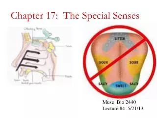
Taste and Olfaction • Receptors for taste and olfaction are chemoreceptors because they respond to chemicals in solution • Thousands of olfactory receptors occupy postage stamp size area in roof of each nasal cavity • Olfactory receptor cells are neurons with olfactory hairs • Long cilia on nasal epithelium that are continually bathed by layer of mucus secreted by underlying glands
Olfaction, cont. • When receptors stimulated by chemicals dissolved in mucus, they transmit impulses along olfactory nerve (1st cranial nerve) to olfactory cortex of brain • Interpretation of odor occurs and “odor snapshot” is made • Olfactory pathways closely tied to limbic system (emotional-visceral part of brain)
Olfaction III • Olfactory impressions are long-lasting and part of one’s memories and emotions • OMG, Ms. Pennington and My Sin® perfume…Beam me up Scotty….I can’t Captain, I ain’t got the power • Olfactory receptors are very sensitive—just a few molecules can activate them • Olfactory neurons adapt very quickly