Download
1 / 34
370 likes | 716 Views
MS3 Medicine Core Conference. Chest Radiology Part 1: The Normal Chest X-Ray. Omar M. Albustami, MD. Pulmonary Disease and Critical Care Medicine Fellow. Brody School of Medicine. East Carolina University. July 06, 2010. Outline. Section A: How to look at a CXR

E N D
MS3 Medicine Core Conference Chest RadiologyPart 1: The Normal Chest X-Ray Omar M. Albustami, MD. Pulmonary Disease and Critical Care Medicine Fellow. Brody School of Medicine. East Carolina University. July 06, 2010
Outline Section A:How to look at a CXR Basic interpretation is easy Technical quality Scanning the PA film How to look at the lateral film. Section B:Localizing lesionsLungs Heart Ground rules that must be applied when interpreting the CXR.
CXR • Powerful investigation mass of info about the cardioresp disease. • Easily available. • Cheap. • Safe. • Value depends on the quality of the clinician viewing the film.
Section A: How to look at a CXRBasic interpretation is easy • Adopt the following procedure: • Name & date. • Technical quality. • Scan & mentally list any abnormalities. Do not stop when you find the first abnormality. • If abnormalities are found, work out where they are. • Mentally describe the abnormality. Which category does it fall into: • Too white. • Too black. • Too large. • In the wrong place.
Technical quality PA AP
Technical quality • Always check the technical quality of any film before interpreting it further. • Examine: • Projection. • Orientation. • Rotation. • Penetration. • Degree of inspiration.
Technical qualityProjection: defined by the direction of the x-ray beam in relation to the pt. Portable (AP or Antero-posterior) PA (Postero-anterior)
Technical qualityOrientation • Check the left/right markings (can be wrong). • Do not assume the heart is always on the left: • Dextrocardia. • The mediastinum can be pushed or pulled to the right by lung pathology.
Technical qualityRotation • Medial ends of the clavicles should be equidistant from the spinous process. • If one clavicle is nearer than the other pt is rotated the lung on that side will appear whiter.
Technical qualityPenetration • Look at the lower part of the cardiac shadow. • The vertebral bodies should only just be visible through the cardiac shadow at this point. • Too clearly visible: film is over penetrated may miss low density lesions. • Cannot see them: under penetrated lung fields will appear falsely white. • When comparing x-rays, the level of penetration should be taken into consideration.
Technical quality Degree of inspiration • Count the number of ribs above the diaphragm. • Midpoint of the right hemidiaphragm should be between 5th -7th ribs anteriorly. • Anterior end of 6th rib • Post end of 10th rib • Poor inspiration will: • make the heart look larger, • give the appearance of basal shadowing & • cause the trachea to appear deviated to the right. should be above the diaphragm.
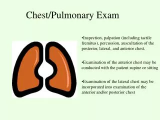
Scanning the PA film • Find a decent viewing box with a functioning light that does not flicker. • Lower the ambient light. • Survey the x-ray from a distance (4 feet) then close up. • Check list: • Lung fields. • Hilum. • Heart. • Rest of mediastinum. • Diaphragms & costophrenic angles. • Trachea. • Bones. • Soft tisuues.
Check list • Lung fields: • Equal transradiancy. • Horizontal fissure: should run from hilum to 6th rib in the axillary line. • Any discrete or generalized shadows. • Hilum: Left should be higher than right (< 1 inch difference): should be concave in shape and have similar density. • Heart: • Shape. • Diameter: <50% transthoracic diameter. • Margins should be sharp. • Dense areas. • Rest of mediastinum: Edge should be clear though some fuzziness is acceptable at the angle between heart & diaphragm, apices & Rt hilum. • Trachea: • Should be central but deviates slightly to the right around the aortic knuckle. • Right side white edge width <2-3 mm on an erect film.
Check list…cont’d. • Diaphragms & costophrenic angles: • Right diaphragm is higher than left (< 1.2 inch difference). • Outline should be smooth. • Costophrenic angles: Well defined & acute. • Bones: • Density. • Compare both sided. • Areas of blackness. • Soft tisuues: • Enlargment. • Gas.
How to look at the lateral film • Name & date. • Step back. • Identify diaphragms: • 1: right hemidiaphragm: can be seen to stretch across the whole thorax & clearly seen passing through the heart border. • 2: left hemidiaphragm: seems to disappear when it reaches the post border of the heart. • Costophrenic angles. • 3: Gastric air bubble.
How to look at the lateral film • Lung fields: • compare appearance in front of & above heart to those behind (equal density). • Discrete lesions. • 4: Retrosternal space: ? Ant mass. • 5: Horizontal fissure: faint white line passing horizontally from the midpoint of the hilum to the ant chest wall. • 6: Oblique fissure: T4/T5 vertebrae, through the hilum, ending at the ant third of the diaphragm. • 7: Hila. • Vertebral bodies: • all same shape & size. • Density: get more translucent (darker) caudally.
Section B:Localizing lesions: Lungs To accurately localize a lesion on CXR, we need to look at both the PA & lateral films. PA film: • Horizontal fissure. • Borders of the lesion: if the lesion is next to a dense (white) structure, the border will be lost silhouette sign. • RML lesion obscures part of the heart border. • RLL lesion obscures the border of the diaphragm.
Localizing lesions: Heart • PA film: • Right heart border up from the diaphragm: • Edge of right atrium. • Above the hilum: SVC. • Left heart border up from the diaphragm: • Left ventricle. • Concavity: left atrial appendage. • At the level of the hilum: pulmonary artery. • Aortic knuckle.
Cardiac Silhouette • R Atrium • R Ventricle 3. Apex of L Ventricle • Superior Vena Cava • Inferior Vena Cava 6. Tricuspid Valve • Pulmonary Valve • Pulmonary Trunk 9. R PA 10. L PA
Lateral film: Heart Ant border: Right ventricle. Post border: Left ventricle.
Location of cardiac valves is best determined on the lateral CXR. • Draw an imaginary line from the apex of the heart to the hilum. • The pulmonic & aortic valves generally sit above this line and the tricuspid & mitral valves sit below.