Download
1 / 9
110 likes | 390 Views
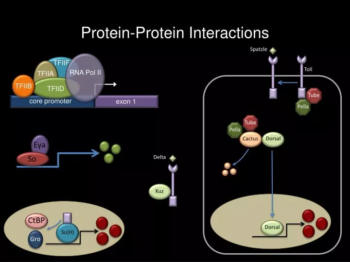
Protein-Protein Interactions. Spatzle. Toll. RNA Pol II. TFIIF. TFIIB. TFIID. Tube. Tube. core promoter. exon 1. Pella. Pella. TFIIA. Dorsal. Dorsal. Cactus. Kuz. Delta. Eya. CtBP. Gro. So. Su(H ). Detecting Protein-Protein Interactions Yeast 2-Hybrid (Y2H) Assays.

E N D
Protein-Protein Interactions Spatzle Toll RNA Pol II TFIIF TFIIB TFIID Tube Tube core promoter exon 1 Pella Pella TFIIA Dorsal Dorsal Cactus Kuz Delta Eya CtBP Gro So Su(H)
Detecting Protein-Protein InteractionsYeast 2-Hybrid (Y2H) Assays - One method for detecting protein-protein interactions is the the yeast 2-hybrid (Y2H) assay. In this assay two proteins are tested for their ability to interact with each other. One protein is fused to the GAL4 DNA binding domain (X-GAL4 BD) while the second protein is fused to the GAL4 activation domain (Y-GAL4 AD). Plasmids containing DNA sequences for these two chimeric proteins along with a plasmid containing a UAS-lacZ reporter construct are simultaneously transformed into yeast cells. Expression of the two fusion proteins is induced and the yeast cells are screened for the presence of the beta-galactosidase enzyme (remember that this enzyme can break down a lactose analog into a blue precipitate). - If the two proteins (X and Y) interact then the GAL4 activation domain will be brought into contact with RNA Pol II which will transcribe the lacZ gene. If this happens then the yeast cells will turn blue. If the two proteins fail to interact then the GAL4 activation domain will not be in the correct position to stimulate RNA Pol II. In this case the yeast cells will remain white. - The Y2H assay can be used in two different contexts. In one case you can test the ability of just two proteins to interact. In another context you screen all of the proteins that are encoded by the genome to interact with one particular protein of interest. In the latter scenario a library of cDNAs is cloned into plasmids containing GAL4 AD. This plasmid library is transformed into populations of yeast cells. These cells are co-transformed with a plasmid containing the gene of interest fused to GAL4-BD. The yeast colonies are screened for putative protein-protein interactions. GAL4-AD Y GAL4-BD Protein X – GAL4 BD UAS-lacZ Protein Y – GAL4 AD GAL4-AD Y RNA Pol II X X GAL4-BD promoter 5X (UAS) lacZ
Co-Immunoprecipitations (co-IP) of Proteins Transfect Cell Lines and Express Proteins of Interest - Another approach to detecting protein-protein interactions is through the use of the co-immunoprecipitation (co-IP) technique. Cells are transfected with plasmids that encode the two proteins that are being tested. The proteins are extracted from the cells and an antibody recognizing one of the two proteins (red) is added to the protein lysate. The antibody-protein complex is purified – this is called the immunoprecipitation (IP) step. The antibody-protein lysate is then run onto a gel and the proteins are transferred to filter paper. Antibodies against both proteins are then added to the filter paper – this is the immunoblot (IB) step. Using sophisticated methods one can determine if the antibodies can detect either of the two proteins. - If the two proteins (red and green) form a complex then two bands will be seen on the filter paper – one representing the red protein and one representing the green protein. The red protein is isolated since it came through the IP step. The green protein is detected in the IB step since it is part of the complex. - If the two proteins do not interact then only one band will show up – the red protein. It was isolated in the IP step. Since the green protein did not interact with the red protein it is not loaded onto the gel. Y Isolate proteins Add antibody against protein X Purify antibody-protein complex Run proteins on a gel Y Y Y Y Y Y Y Transfer proteins to filter Add antibodies against protein X and Y Y Y Y Y Y Y Y
Detecting in vivo Protein-Protein Interactions • The split YFP assay is an in vivo assay that can be used to determine if two proteins physically interact in a cell. YFP is a variant of GFP that emits light in a different wavelength than GFP. It can be split into two halves – neither half will glow yellow. However, if both halves are allowed to interact then YFP is reconstructed and it will glow yellow. • In our example protein A is fused to one half of YFP and protein B is fused to the other half of YFP. Both proteins are then expressed in a cell or a tissue. If the two proteins interact (above right) then the two halves of YFP can interact thereby reconstituting the full-length YFP protein (which glows yellow). • If the two proteins do not interact then the two halves of YFP will not come together – and the cell will not glow at all. B A Question: Do proteins A and B physically interact? YFP A B
Detection of Proteins: the Western Blot • The Western Blot is a method for determining if a particular protein is present within a cell population, a tissue or an organ. It can be used to compare wild type and mutant samples as well as samples from different developmental time points. • Methods exist for isolating proteins from cells and tissues (the protein slurry is called a lysate). The entire protein lysate is loaded onto a polyacrylamide gel and separated by size (using an electric current). The protein lysate is then transferred to a nitrocellulose membrane to which they are affixed using a cross-linking reagent. Chemiluminescent labeled antibodies are incubated with the filter and used to detect the protein of interest. • There are chemicals that can detect all proteins without any regard to specificity. It is also possible to use labeled antibodies to detect specific proteins. In the former case all proteins within a lysate will be visualized. In the latter case only a subset of proteins will be detected. • Protein detection starts with a primary antibody that recognizes the protein of interest. A secondary antibody that is labeled with a chemiluminescent tag is then used to bind to the primary antibody.
Detection of Proteins: the Western Blot • The Western Blot is a method for determining if a particular protein is present within a cell population, a tissue or an organ. It can be used to compare wild type and mutant samples as well as samples from different developmental time points. • Methods exist for isolating proteins from cells and tissues (the protein slurry is called a lysate). The entire protein lysate is loaded onto a polyacrylamide gel and separated by size (using an electric current). The protein lysate is then transferred to a nitrocellulose membrane to which they are affixed using a cross-linking reagent. Chemiluminescent labeled antibodies are incubated with the filter and used to detect the protein of interest. • There are chemicals that can detect all proteins without any regard to specificity. It is also possible to use labeled antibodies to detect specific proteins. In the former case all proteins within a lysate will be visualized. In the latter case only a subset of proteins will be detected. • Protein detection starts with a primary antibody that recognizes the protein of interest. A secondary antibody that is labeled with a chemiluminescent tag is then used to bind to the primary antibody.
Detection of Proteins: the Western Blot • The Western Blot is a method for determining if a particular protein is present within a cell population, a tissue or an organ. It can be used to compare wild type and mutant samples as well as samples from different developmental time points. • Methods exist for isolating proteins from cells and tissues (the protein slurry is called a lysate). The entire protein lysate is loaded onto a polyacrylamide gel and separated by size (using an electric current). The protein lysate is then transferred to a nitrocellulose membrane to which they are affixed using a cross-linking reagent. Chemiluminescent labeled antibodies are incubated with the filter and used to detect the protein of interest. • There are chemicals that can detect all proteins without any regard to specificity. It is also possible to use labeled antibodies to detect specific proteins. In the former case all proteins within a lysate will be visualized. In the latter case only a subset of proteins will be detected. • Protein detection starts with a primary antibody that recognizes the protein of interest. A secondary antibody that is labeled with a chemiluminescent tag is then used to bind to the primary antibody.
Molecular Biology Study Questions - What types of interactions are detected by the Y2H assay? - Why do you fuse the GAL4 activation and DNA binding domains to the proteins that you are testing for interactions? - In the Y2H assay how do you know if the the two proteins of interest physically interact? - What types of interactions are detected by the co-IP assay? - What is difference between the IP and the IB steps? - Why do you add only one antibody during the IP step? - Why do you add both antibodies during the IB step? - In a co-IP assay, how do you know if two proteins physically interact? - In the Split – YFP assay how do you know if two proteins interact within a cell? - What is the purpose of the Western blot? - Which assay (Y2H, Co-IP, Western Blot or Split-YFP) detects in vivo protein interactions?
Preview of Upcoming Lecture Topics to be Covered Next Time Anterior-Posterior Patterning Maternal Effect, Gap, Pair-Rule, Segment Polarity, and Homeotic Genes 3`UTRs, RNA localization, Microtubules and Motors Diffusion Gradients (proteins and mRNA transcripts) Transcriptional Repressors and Body Plan Formation • Textbook Chapter • Chapter 21 pg. 751-772 Weekly Article(s): for next week “The Evolution of Color Vision” “Ingenious” “Gene Therapy”