Download
1 / 72
730 likes | 957 Views
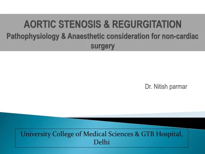
AORTIC STENOSIS & REGURGITATION Pathophysiology & Anaesthetic consideration for non-cardiac surgery. Dr. Nitish parmar. University College of Medical Sciences & GTB Hospital, Delhi. Objectives. Definition Etiology Pathophysiology Preoperative evaluation Anaesthetic management.

E N D
AORTIC STENOSIS & REGURGITATIONPathophysiology & Anaesthetic consideration for non-cardiac surgery Dr. Nitishparmar University College of Medical Sciences & GTB Hospital, Delhi
Objectives Definition Etiology Pathophysiology Preoperative evaluation Anaesthetic management AS/AR
Normal P-V loop esv edv
Laplace law Wall tension = P x R / 2h P= pressure R = radius h= wall thickness Increase wall tension stimulates concentric hypertrophy of LV
Aortic stenosis (AS) • Aortic stenosis refers to obstruction of flow at the level of the aortic valve • Restricted systolic opening of the valve, with a mean transvalvular pressure gradient of at least 10 mm Hg. • Normal aortic valve area is 2.5-3.5cm² • Haemodynamically significant obstruction occurs at valve area of < 1 cm2
Pathophysiology Chronic pressure overload Peak systolic wall stress Parallel sarcomere replication Concentric hypertrophy ( no increase in the LV size )
AORTIC STENOSIS LV outflow obst. ↑LV systolic P ↑ LVET ↑ LV diastolic P ↓ AoP ↑ LV mass ↑ Myocardial O2 consumption ↓ Diastolic time Myocardial ischemia LV Dysfunction LV Failure
Contd… • Increased diastolic stiffness magnifies the importance of atrial systole to ventricular filling • Hypertrophied ventricle is highly sensitive to ischemia • Increase myocardial oxygen demand • Decreased coronary perfusion gradient • Circulatory abnormalities in myocardium • Decreased capillary density • Abnormal thickening of coronary arterioles
Preoperative evaluation angina dysopnea syncope
Angina Myocardial O2 mismatch • ↑Demand • due ventricular hypertrophy • ↓Supply • ↑ LVEDP • Hypotension • Abnormal coronary circulation • ↓capillary density • Thickened arterioles
Dyspnea Systolic and diastolic dysfunction of left ventricle pulm capillary hydrostatic pressure transudation of fluid into interstitial space lung compliance WOB dyspnea
Syncope • fall in CO d/t arrythmia fixed CO state • Other symptoms • Symptoms of LVF occur only in the advanced stages of the disease • When AS and MS coexist volume produced by MS gradient across the AV masking of clinical findings by AS
signs • Pulse: small volume, pulsusparvus et tardus • BP – normal or low • PP – low, due to fall in SBP when SV in late stages
Signs • Cardiac apex – heaving • S1 – normal / soft • S2 – paradoxical split in late stages • Palpable S4 • Aortic ejection click • Ejection mid systolic murmur
Ecg LVH LV- strain pattern
CXRProminent ascending aortarounding of left ventricular apexcalcified valves
Echo Asessment of severity However when cardiac output is low severe stenosis may be present with lower transvalvular pressure gardient and lower jet velocity
Non cardiac surgery elective emergency Proceed with medical optimization and high risk severe Mild to moderate GA/spinal /epidural
SEVERE asymptomatic symptomatic Risk of surgery Risk for AVR Low/mod High Low High Risk for AVR High Low Consider valvuloplasty High risk stratification Proceed AVR Consider valvuloplasty High risk stratification
Monitoring • Standard non-invasive • ECG : 5 lead including lead II & V5 • HR • NIBP • Pulse-oxymetry • Capnograph • Temperature • Apply defibrillator pads beforehand
Monitoring • Invasive monitoring • IBP • CVP/PAC ?? • Echocardiography (TEE)
Premedication Aim • To decrease anxiety & any associated likelihood of adversecirculatory responses produced by tachycardia
Post-operative management Majority of the cardiac events in non cardiac surgery occur in postoperative period Monitoring Oxygen Pain relief: multimodal including neuroaxialopioids Intravenous fluids
Regional anesthesia • Technique should be applied cautiously • Mild to moderate aortic stenosis: can be used • Severe : contraindicated • Epidural anesthesia is preferable to spinal anesthesia • Spinal with opioid • Caution with anticoagulants
Prosthetic valves Prosthetic valves • Mechanical • Greater durability • Needs life long anticoagulation • More complications • Preferred in younger patients (<65) • Bioprosthetic • Less durable • No need for anticoagulation • Lesser complications • Preferred in older patients who donot need anticoagulation
Complications • Valve thrombosis • Systemic embolization • Structural failure • Hemolysis • Paravalvular leak • Endocarditis
Endocarditis prophylaxis • Dental procedures • Procedures involving incision of respiratory mucosa (adenoidectomy, tonsillectomy) • Incision on infected skin, musculoskeletal tissue • Cystoscopy if results of urine culture not known
Anticoagulation Discontinue warfarin at least 5 day before Sx Bridge with SC LMWH or IV unfractionated heparin (36 hrs later) Asses INR 1-2 days before Sx If >1.5 1-2 mg oral vit K
Anticoagulation Patient receiving SC LMWH-stop 24 hrs prior Patient receiving UF heparin-stop 4 hrs prior Post operative Emergent procedure: treat with 2.5-5.0 mg IV vitamin K For faster reversal: FFP • Major Sx • Minor Sx • Begin after 48-72 hrs • Begin after 24 hrs
Anticoagulation Start the patient back on oral warfarin Monitor PTT and aPTT on daily basis Attain INR of 2.5-3.0 for 2-3 days Stop heparin and continue warfarin
Neuraxial anesthesia • Neuraxial block should be delayed for 12 hrs (prophylactic) and 24 hrs (therapeutic) after last dose of LMWH • Removal of epidural catheter should take place 12 hrs after last dose • Subsequent dosing delayed for 2 hrs
Preoperative • Echo • Ejection fraction • Normal functioning of valve • Paravalvular leaks • Vegetations • Clots • In a patients with prosthetic valves rest of the anesthetic concerns are similar to a regular non VHD patient
Aortic regurgitation incompetence of the aortic valve, in which a portion of the left ventricular forward stroke volume returns to the chamber during diastole.
Etiology In approximately two third of the patients with valvular AR, the disease is rheumatic in origin • Acute • IE • Trauma • Aortic dissection • Chronic • Abnormalities of • AV (congenital bicuspid valve) • Aortic root – syphilis, cystic medial necrosis • Marfan’s syndrome • Rheumatic arthritis
Pathophysiology LV cannot dilate sufficiently Effective SV Sudden in LVEDP Transmitted to pulm circ. Acute pulm congestion
Contd.. Chronic overload ↑LVEDV ↓ Series replication of sarcomere ↓ Chamber enlargement ↓ Eccentric hypertrophy ↑ wall stress ↓ Concentric hypertrophy cardiomegaly (mild)