Download
1 / 10
100 likes | 216 Views
Noninvasive In V ivo Imaging of Tissue Pathology. Stavros G. Demos, Ph.D. Physicist, LLNL . In Vivo Histopathology: The “Microscope” Healthcare has been waiting for…. . Normal Esophagus. Barrett’s Esophagus. Autofluorescence Capturing UV Microscope.

E N D
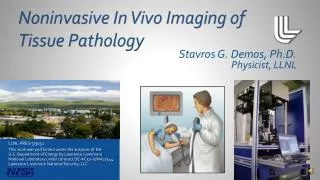
Noninvasive In Vivo Imaging of Tissue Pathology Stavros G. Demos, Ph.D. Physicist, LLNL
In Vivo Histopathology: The “Microscope” Healthcare has been waiting for…. Normal Esophagus Barrett’s Esophagus AutofluorescenceCapturing UV Microscope • In vivo imaging of tissues microstructure • Instantaneous diagnostic decision-making • Non-invasive • Reduces overall time spent in surgery • Eliminates anxiety in waiting for diagnosis • Reduces overall costs • Greatly improves healthcare
Tissue Biopsy is the Gold Standard but….. Source: murrasaca.com • Tissue sample is removed from the patient, sent to the lab, sliced into sections, mounted on slides, stained, and examined under microscope • Invasive • Results in multiple and repetitive surgeries • Imprecise • Random sampling error often misses pre-cancerous cells • Feedback takes extended amounts of time, (typically days) • Costs are extremely high
The Healthcare system must Advance the Century-old Biopsy Method with…. Source: www.gastroarkansas.com Endomicroscope systems • Instantaneous/real-time diagnostic info. for surgeon • Early, instant, accurate detection of disease (cancer, etc.) • Imaging helps guide surgical intervention • Noninvasive • Reduces repetitive surgeries • Precise cellular level images • Random sampling error is not a factor • Feedback is instantaneous • Drastically reduces overall costs
Emerging competing In Vivo Microscope Technology has Significant Limitations Mauna Kea’s Confocal Laser Endomicroscopy product: *optical sectioning *fluorescein Source: maunakeatech.com Current Problem: conventional endomicroscope systems yield blurry images Current Market Solutions: 1) Optical sectioning ⇨ trade-off: poor light collection efficiency 2) Contrast agents⇨ trade-offs: long preparation time and expense
Patented LLNL Solution: Microscope Design that captures Autofluorescence under UV Excitation UV Excitation eliminates need for Optical Sectioning: a) Limits photon penetration depth b) Allows high throughput designs w/ optimal signal collection Autofluorescenceeliminates need for Contrast Agents/Staining: (Intrinsic UV excited fluorophores) a) Act as “natural stains” Pulsed Illumination yields resolution suitable for tissue diagnosis:a) Provides diffraction limited spatial resolution b) Eliminates motion artifacts in vivo
Physicians now can have Access to In VivoImages of Tissue Pathology Epithelium of Stomach AF capturing UV Microscope: Real Time Features seen are comparable to Gold Standard, BUT AF capturing UV microscope provides greater speed, ease, & cost efficiency Gold Standard: Tissue biopsy H&E Stain Visualization at the cellular level Details of epithelial microstructure Visualization of the margins Targeted biopsies High resolution images (spatial resolution ~ 1 µm)
Real-Time Imaging Remains a Versatile Technology Heeryae.com
Technology is Ready to Move and Excel Source: businessinsider.com Available tabletop prototype Tested microendoscope concept and design parameters (ex vivo) Diagnostic studies: human esophageal tissues and animal tissues (ex vivo) Animal imaging and stem cell localization/targeting Future Development: Proof of Principle in Humans (in vivo)
Contact Information GenaroMempin mempin1@llnl.gov