Download

1 / 30
300 likes | 591 Views
Functional Human Physiology for the Exercise and Sport Sciences Synaptic Transmission and Neural Integration. Jennifer L. Doherty, MS, ATC Department of Health, Physical Education, and Recreation Florida International University. Types of synapses. Electrical Synapses

E N D
Functional Human Physiologyfor the Exercise and Sport Sciences Synaptic Transmission and Neural Integration Jennifer L. Doherty, MS, ATC Department of Health, Physical Education, and Recreation Florida International University
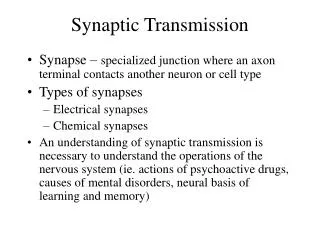
Types of synapses • Electrical Synapses • Nerve impulse transmission directly from one neuron to another through membranes connected by gap junctions • No neurotransmitters • Chemical Synapses • Nerve impulse is transmitted from one neuron to another across a synapse • A region of functional contact (not actual contact) with another neuron • Specialized to release and receive neurotransmitters
Chemical Synapses – Functional Anatomy • Neuron-to-Neuron Synapses • Close junction between the axon terminal of one neuron and the plasma membrane of another neuron • Presynaptic Neuron • At a synapse, a neuron that transmits signals to a second neuron • Communicates with a post-synaptic neuron through the axon
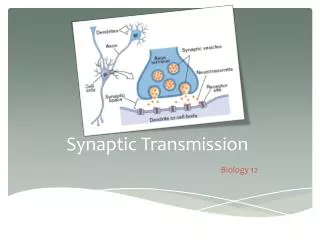
Chemical Synapses – Functional Anatomy • Synaptic Knob (Bouton) • The rounded terminal end of the axon on a presynaptic neuron • Many synaptic knobs of many axons may terminate on the cell body or dendrites of postsynaptic neurons. • Contains many mitochondria • Contains Synaptic Vesicles • Store neurotransmitters
Chemical Synapses – Functional Anatomy • Synaptic Vesicles • Release neurotransmitters to diffuse across the synaptic cleft • Neurotransmitters attach to receptors on the post-synaptic neuron cell membrane • Synaptic cleft • Narrow, fluid-filled space between presynaptic and post-synaptic neurons • 30 - 50 nm or 1 millionth of an inch wide • No direct contact between neurons.
Chemical Synapses – Functional Anatomy • Post-synaptic neuron • At a synapse, the neuron that receives signals from another neuron • Post-synaptic membrane • Contains neurotransmitter receptors • Specialized protein receptors that react with (or receive) a specified neurotransmitter
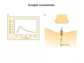
Signal Transduction – Nerve Impulses (Wave of Action Potentials) • Presynaptic Neuron Signal Transduction • A wave of action potentials are propagated along the cell membrane of the presynaptic axon • The wave of action potentials arrive at the synaptic knob. • Ca++ voltage-gated channels in the synaptic knob open in response to depolarization of the cell membrane • Ca++ ions from the extracellular fluid enter the synaptic knob
Signal Transduction – Nerve Impulses (Wave of Action Potentials) • Increased amounts of intracellular Ca++ ions • Causes the synaptic vesicles to move toward and fuse with the membrane of the synaptic knob • The synaptic vesicles release neurotransmitters into the synaptic cleft via exocytosis • Unless another action potential is present… • The Ca++ voltage-gated channels close • Ca++ is actively transported out of the synaptic knob.
Signal Transduction – Nerve Impulses (Wave of Action Potentials) • Synaptic Cleft • Neurotransmitters diffuse across the synaptic cleft • Transmission of a nerve impulse across the synaptic cleft can only occur in one direction • Neurotransmitters bind to specific protein receptors on the post-synaptic membrane • Produces a response on the post-synaptic neuron • Neurotransmitters are quickly removed from the protein receptors by: • Degradation by specific enzymes, • Diffusing away from the synapse, or • Reuptake by neuroglial cells or the presynaptic neuron
Signal Transduction – Nerve Impulses (Wave of Action Potentials) • Post-Synaptic Signal Transduction • Neurotransmitters bind to specific protein receptors on the post-synaptic membrane • Ion channels on the post-synaptic membrane open • Results in a change of the membrane potential of the post-synaptic neuron • Depolarization occurs • Results in a wave of action potentials that is propagated along the cell membrane of the post-synaptic neuron
Signal Transduction – Nerve Impulses (Wave of Action Potentials) • Rarely is only one neuron responsible for producing an action potential on the cell membrane of another neuron • Several neurons must produce enough graded potentials to reach the “threshold” for generating an action potential • Two determinants of signal transduction: • The type of neurotransmitter, and • The receptor proteins • Result is either • Excitation (depolarization), or • Inhibition (hyperpolarization)
Excitatory Synapses • Bring the membrane potential closer to the threshold for generating an action potential • Excitatory neurotransmitters depolarize the post-synaptic membrane • This is called an Excitatory Postsynaptic Potential (EPSP) • Opens Na++ voltage-gated channels on the post-synaptic membrane allowing Na++ ions to enter the cell • Fast response occurs via Ionotropic receptors • Channel-linked receptors • Slow response occurs via Metabotropic receptors • G-protein linked receptors
Inhibitory Synapses • Bring the membrane potential away from the threshold for generating an action potential • Inhibitory neurotransmitters hyperpolarize or stabilize the post-synaptic membrane • This is called an Inhibitory Postsynaptic Potential (IPSP) • Opens K+ voltage-gated channels on the post-synaptic membrane allowing K+ ions to leave the cell
Neural Integration • The net effect of EPSPs and IPSPs on the post-synaptic membrane will determine if the net effect is excitatory or inhibitory. • If the net effect is more excitatory than inhibitory, an action potential will be generated on the post-synaptic membrane and impulse transduction will occur • The opposite is also true, a net inhibitory effect will not produce an action potential.
Neurotransmitters • Structure • Chemical compounds • Over 30 different types of neurotransmitters • Major categories include: • Choline derivatives • Biogenic amines • Amino acids • Neuropeptides • Synthesis • Neurotransmitters are synthesized in the cytoplasm of the nerve cell body or the synaptic knob • Neurotransmitters are stored in the synaptic vesicles • More than one neurotransmitter may be produced by a neuron
Acetylcholine (ACh) • Choline derivative neurotransmitter • Found at the neuromuscular junction predominantly • Responsible for stimulating muscles to contract • Acetylcholinesterase • Enzyme that degrades ACh into acetate and choline • Acetylcholine Receptors • Nicotinic cholinergic receptors • Excitatory • Found on skeletal muscles (somatic nervous system) and in neurons in the autonomic nervous system • Muscarinic cholinergic receptors • Either excitatory or inhibitory • Found in the central nervous system
Biogenic Amines – Neurotransmitters derived from Amino Acids • Catecholamines • Includes the neurotransmitters: • Norepinephrine (NorE) • Epinephrine (Epi) • Dopamine • Important in many of the motor functions of the autonomic nervous system • Serotonin • Found in brainstem • Regulates sleep and emotions • Histamine • Found in hypothalamus • Better known for release from tissue cells in inflammatory response
Biogenic Amines – Neurotransmitters derived from Amino Acids • Adrenergic receptors • Alpha adrenergic • Norepinephrine mostly, but binds to both • Beta adrenergic • Epinephrine mostly, but binds to both • Degradation enzymes: • Monoamine oxidase (MAO) • Catechol-O-methyltransferase (COMT)
Amino Acid Neurotransmitters • Most abundant class of neurotransmitters • Function only in the central nervous system • Excitatory neurotransmitters • Glutamate • Aspartate • Inhibitory neurotransmitters • Glycine • Gamma-aminobutyric acid (GABA)
Neuropeptides • Short chains of amino acids that function as neuromodulators • A neuromodulator is a substance that alters the response of a neuron to a neurotransmitter, or it blocks the release of a certain neurotransmitter. • Neuropeptides are more classically known as hormones • Examples: • TRH = regulates the release of TSH • Substance P = decreases gastrointestinal motility • Vasopressin = regulates urine output by the kidneys • Endogenous opiates = analgesic effect, euphoric response • Enkaphalins • Endorphis