Download
1 / 19
200 likes | 437 Views
Lower GI Disease. Lori F Gentile. Lower GI Disease. Diverticular disease Bowel Obstruction Appendicitis Colon Cancer Inflammatory Bowel Disease Volvulus Olgilvie’s Syndrome Lower GI Bleed Ischemic Bowel Disease. Obstruction. LGI-distal to the Ligament of Trietz

E N D
Lower GI Disease Lori F Gentile
Lower GI Disease • Diverticular disease • Bowel Obstruction • Appendicitis • Colon Cancer • Inflammatory Bowel Disease • Volvulus • Olgilvie’s Syndrome • Lower GI Bleed • Ischemic Bowel Disease
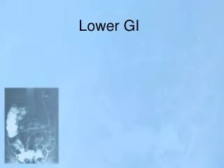
Obstruction • LGI-distal to the Ligament of Trietz • Ileus = obstruction 2/2 dysfunctional motility of bowel • Mechanical obstruction = 85% SB, 15% large bowel • Simple obstruction, closed loop obstruction, Strangulation • Most Common Cause • Pt with previous surgery Small bowel – Adhesions Large Bowel – Cancer • Pt without previous surgery Small bowel – Hernia Large Bowel - Cancer
SBO: Etiology • Adhesion • Hernia • Tumor • Abscess • Hematoma • Annular pancreas • SMA syndrome • Congenital lesions • Gallstone ileus • Intussusception • Foreign body (bezoars, worms, etc) • Meconium ileus • Malrotation
Colonic Obstruction: Etiology • Cancer #1 (60%) • Volvulus (sigmoid > cecum) • Adhesions • Hernia • UC • Diverticulitis • Congenital lesions • Fecal impaction • Adynamic ileus • Hirschsprung’s • Meconium ileus • Foreign body
History & DDx • Proximal obstruction: early bilious vomiting, +/- flatus/BM • Distal obstruction: obstipation, distension, vomiting feculent material (2/2 bacterial overgrowth of SB contents) • Pain w/obstruction: begins as cramping pain, changes to continuous severe pain w/strangulation & peritonitis • Bowel Movements- Cabliber, Blood, Pain • PMHx: remember to ask about cardiac history (arrhythmias, prior MI, Afib - think about intestinal ischemia), IBD, gallstones, cancer • PSHx: remember to ask about ostomy output • Meds: narcotics (ileus), antipsychotics (ileus), diuretics (hypoK a/w ileus) • ROS: recent weight loss (CA)
PE • Start with ABCs • Look for surgical scars • Bowel sounds • Distention-> tympany to percussion • Localized tenderness • Look for hernias/masses • Do a rectal exam
Labs • WBC (nml in uncomplicated SBO) • CBC (anemia w/CA) • BMP (hypoK) • Alkalosis (a/w proximal obstruction) • Acidosis (a/w bowel infarction) • Lactic acid- may be indication of bowel ischemia
Studies • Upright CXR/KUB: look for free air • Flat and upright/left lateral decubitus: look for dilated bowel loops, air-fluid levels • Note: if cecal diameter >12cm, there is a risk of perforation. At 12-14cm, the wall tension > perfusion pressure, increasing risk of necrosis • Barium enema • UGI series w/SB follow-through • CT scan- with PO/IV contrast
SBO: Management • “Bowel Rest” - NPO, NGT, Foley, IVF-Cures >80% SBOs • Electrolyte replacement • “Don’t let the sun set on a (complete) SBO” Complete bowel obstruction w/concern for strangulation/perforation requires immediate operative intervention (resuscitate first) • Indications for Surgery -> Failure to resolve, progressing pain, peritoneal signs, fever, increasing WBCs
A 72-year-old woman presented with a 2-day history of abdominal pain associated with nausea and vomiting Dedouit F and Otal P. N Engl J Med 2008;358:1381
A 48-year-old healthy woman presented with anorexia of 2 days' duration and abdominal pain in the right lower quadrant Liu K and Lin B. N Engl J Med 2007;356:1152
Colon Cancer • Asymptomatic – Screening Colonoscopy @ age 50 • Adenoma->TVA (50% harbor cancer) • Sessile, high grade dysplasia increase cx risk • Polypectomy for pathology, adequate for T1 if margins clear • Symptoms – abdominal pain, anemia, constipation, bleeding, weight loss • Sigmoid colon-most common site of primary, constipation • Right colon cancer – anemia, asymptomatic • Work-up - staging • CT C/A/P, CEA • If rectal mass-> EUS
Case • 76 year old man, mass in LLQ, gradual growth, intermittent abdominal pain • Last BM 3 days ago, Nausea, Vomiting • Weight loss • Gradually narrowing caliber stools
Case • Imaging: air fluid levels (obstruction) • “Apple core” lesion in colon • Dx: colon CA • Tx: NPO, NGT, lytes • Staging/monitoring: • Colonoscopy • CEA • Chest CT • Neoadjuvant therapy, Resection • Diverting ostomy http://allbleedingstops.blogspot.com
Diverticular Disease • Herniation of mucosa through colon call at points where arteries enter, increased intra-luminal pressure • 80% Left side, sigmoid colon • Diverticulitis – left • Bleeding – right Diverticulitis- infection/inflammation of colonic wall • Sx- LLQ pain, tenderness, fevers, leukocytosis, emesis, diarrheah • Work-up – CT scan Hinchey Classification – Stage 1-4
Diverticular Disease Treatment : Uncomplicated – Bowel rest, Bactrim/Flagyl (PO or IV) • Increase fiber in diet, stool softeners • Consider elective surgery if second attack occurs (50% chance of recurrence) Complicated – obstruction, fluctuant mass, abscess, peritonitis, fistula, sepsis, Hinchey 3,4 • Abscess-percutaneous drainage, abx • Peritonitis – OR->Hartmann’s procedure Needs colonoscopy in 6-8 weeks when sx resolve – r/o cancer, other diseases
Case • A previously healthy 45-year-old man presents with severe lower abdominal pain on the left side, which started 36 hours earlier. He has noticed mild, periodic discomfort in this region before but has not sought medical treatment. He reports nausea, anorexia, and vomiting associated with any oral intake. On physical examination, his temperature is 38.5°C and his heart rate is 110 beats per minute. He has abdominal tenderness on the left side without peritoneal signs. CT scan shows Hinchey 2 with 4 cm peri-rectal contained abscess. How should his case be managed?
Case • Complete H&P • Admit – pt unable to hydrate himself • NPO, IVFs, IV abx • Percutaneous Drainage of Abscess • As pt improves, ADAT, convert to PO abx • Colonoscopy 6 weeks after discharge • Surgery referral should pt have recurrent diverticulitis