Download
1 / 26
270 likes | 534 Views
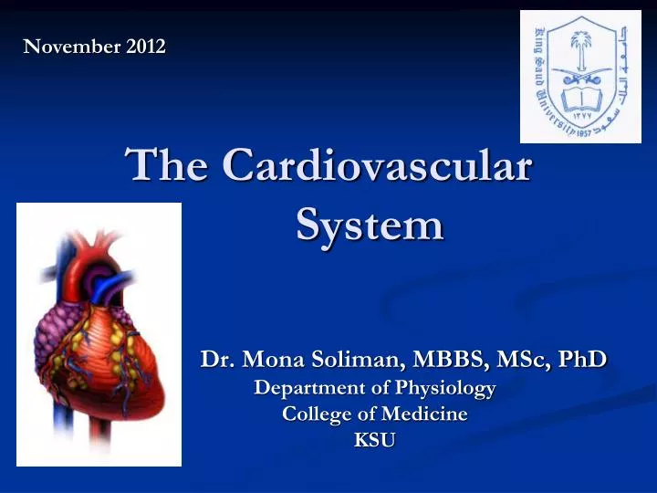
November 2012. The Cardiovascular System. Dr. Mona Soliman, MBBS, MSc, PhD Department of Physiology College of Medicine KSU. Structure of the Heart. Structure of the Heart. The Atria Thin walled Receives blood from: the systemic circulation (right atrium)

E N D
November 2012 The Cardiovascular System Dr. Mona Soliman, MBBS, MSc, PhD Department of Physiology College of Medicine KSU
Structure of the Heart The Atria • Thin walled • Receives blood from: • the systemic circulation (right atrium) • the pulmonary circulation (left atrium) • Open into the ventricles via the: Atrioventricular valves (AV valves)
Structure of the Heart The Ventricles • Thick muscular walled (why?) • Pump blood into: • Pulmonary trunk (right ventricle) • Aorta (left ventricle) • A fibrous tissue ring separate the atria from the ventricles (importance: electrical activity, AV valve)
The Valves of the HeartThe Atrioventricular Valves • The Tricuspid Valve… between the right atrium and the right ventricle, 3 cusps • The Mitral Valve (bicuspid valve) … between the left atrium and the left ventricle, 2 cusps
The Valves of the HeartThe Atrioventricular Valves • Prevent back flow of blood from the ventricles to the atria • Held by chordae tendineae to papillary muscle • Contraction of papillary muscle…
The Valves of the HeartThe Semilunar Valves • Located at the origin of the pulmonary artery and aorta • Open during ventricular contraction…why? • Close during ventricular relaxation…why? • The Aortic Valve • The Pulmonary Valve
Cardiac Muscle cell • Striated • Contain actin and myocin filaments arranged in sarcomeres…contract by sliding mechanism • Branch and interconnect
Cardiac Muscle cell • Gap junctions • Trans-membrane channel proteins, connecting the cytoplasm of the cells • Allow spreading of the action potential from one fiber to another • Allow cardiac muscle to function as a syncytium“all or none law”: stimulation of a single muscle fiber results in contraction of all the muscle fibers • Intercalated discs
Electrical Activity of the Heart • Automaticity: capable of originating action potential
Myocardial action potential • Resting membrane potential in myocardial cells -90 mV Stimulation of myocardial cell Myocardial action potential
Conduction of Impulses • The sinoatrial node (SA node): • Located in the right atrium • Pacemaker of the heart • Is capable of originating action potentials • Highest frequency • The atrioventricular (AV) node • Located at the junction of the atria and the ventricles • Delay in the conduction of impulses…why?
Conduction of Impulses • The atrioventricular (AV) bundle (Bundle of His) • The right and left bundle branches • Purkinje fibers • Spread within the muscle of the ventricular walls • Highest speed of conduction
Contractility • Contractility is the ability of cardiac muscle to convert chemical energy into mechanical work
Contractility Depolarization of myocardial cell Opening of Ca2+ channels Ca2+ increase in the cytoplasm Ca2+ binds to troponin Contraction
Contractility Repolarization of myocardial cell Ca2+ OUT Ca2+ decrease in the cytoplasm Relaxation
Contractility • Absolute refractory period • Cardiac muscle cannot be excited while it is contracting … benefit? • Long ARP • Time: depolarization & 2/3 of repolarization • Relative refractory period • Time: last 1/3 repolarization • Strong stimulus can give rise to contraction