Download
1 / 31
310 likes | 442 Views
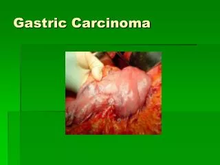
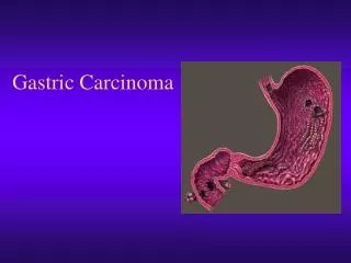
Characteristics of submucosal gastric carcinoma with lymph node metastatic disease. H J Son, S Y Song,1 S Kim,3 J H Noh,2 T S Sohn,2 D S Kim1 & J C Rhee. Presented by intern 張家維. Histopathology 2005, 46, 158–165. Introduction. 2nd most common cause of cancer deaths

E N D
Characteristics of submucosal gastric carcinoma with lymph node metastatic disease H J Son, S Y Song,1 S Kim,3 J H Noh,2 T S Sohn,2 D S Kim1 & J C Rhee Presented by intern張家維 Histopathology 2005, 46, 158–165.
Introduction • 2nd most common cause of cancer deaths • East Asia and south America • Korea and Japan early detection • Early Gastric Carcinoma Mucosa or Submucosa • 5-year survival rate • Lymph node metastasis prognosis, submucosal invasion
Introduction • Depth of submucosal layer viaries • The depth of submucosal gastric carcinoma (SMGC) • Lymph node metastasis macroscopic appearance, location, size, tumor area, differentiation, invasion depth, submucosal vascularity, fibrosis near the tumor area • Age- and Sex- matched
Materials and methods • Sampling: 248 patients SMGC • Surgical resection • Samsung Medical Centre (Seoul, Korea) • 1995/Jan.~ 2002/Oct. • Total 917 patients SMGC 124 LN metastasis (13.5%) • Anticancer therapy X • Evidence of metastatic disease X
Materials and methods • 124 SMGC with LN metastasis • 124 SMGC without LN metastasis • Age- and Sex- matched • Specimens routinely examined • 10% formalin • Embedded in paraffin • H & E stained
Materials and methods • Macroscopic: Japanese Endoscopic Society Classification Elevated, Depressed, Flat • Microscopic: Lauren classification
Materials and methods • Depth of submucosal invasion 1.Ocular lens scale, distance between the lower edge of the muscularis mucosa and the deepest invading front of the tumor cells 2.sm3 method: sm1, sm2, sm3 3.sm2 method: smi , sme
Materials and methods • Tumor size Longest dimension of the tumor area • Tumor area Longest dimension X its prependicular counterpart • Tumor vessels vessels with a smooth muscle coat in the submucosa
Materials and methods • Statistical analysis: Log linear model, McNemar’s test, A paired t-test, Wilcoxon’s signed rank test • P-value < 0.05 statistically significant • SAS, version 6.12
ResultsPathological parameters and lymphnode metastatic disease • 124 SMGC with lymph node metastasis 69 males (55.6%), 55 females (44.4%) 31~83 y/o, the mean age 56.3 y/o • The main locations of the tumors 60 lower, 61 middle, 3 upper 1/3 • Tumor size 5~125 mm, the mean = 45 mm • Tumor area 0.8~105.0 cm2, the mean = 17.1 cm2
ResultsPathological parameters and lymphnode metastatic disease • The gross types of the tumors 35 elevated (28.2%), 85 depressed (68.5%), 4 flat (3.2%) • The histological differentiations (Lauren’s) 53 intestinal (42.7%), 65 diffuse (52.8), 6 mixed (4.8%) • 111 N1 (89.5%), 13 N2 (10.5%)
ResultsPathological parameters and lymphnode metastatic disease • Significantly associated with node-positive SMGC 1. presence of lymphatic tumor emboli 2. a larger tumor area 3. a larger tumor size 4. a non-flat gross type 5. an increased vascularity • No significant relationship 1. location 2. Lauren classification 3. tumor related fibrosis
ResultsDepth-related parameters and lymph node metastatic disease • Ocular scale-measured depth 1. proved to have a significant correlation with node-positive SMGC 2. superficial invasive, deeply invasive (2mm) • The sm3 method not well correlated • The sm2 method not well correlated
ResultsMultivariate analyses for possible indicators of LN metastatic disease • Multivariate logistic regression analysis location, gross type, Lauren’s classification, lymphatic tumor emboli, increased vascularity, tumor-related fibrosis, tumor size, depth (sm2 method) • The incidence of lymph node metastatic disease increased in the presence of lymphatic tumor emboli and in the tumors that invaded more than half of the submucosal layer
Discussion • EGC, the “early” horrible disaster curable disease early diagnosis and treatment programs • The term of EGC has 2 innate defect 1. lymph node metastatic diseases 2. discriminate submucosal tumor call invasion • 5-year survival rate 93~99% for node-negative EGC 73~90% for node-positive EGC 90~100% for intramucosal confinement 73~90% for submucosal invasion
Discussion • The treatment now for EGC conducting minimally invasive surgical procedures endoscopic mucosal resection, laparoscopic partial resection need careful and intensively subclassification • Remove all metastatic lymph nodes ? chance of a cure↓ • Factors related to lymph node metastatic disease
Discussion • SMGC lymph node metastasis rate 10~25% • 917 SMGC in this tiral 13.5% LN metastasis • The parameters related to LN metastasis lymphatic tumor emboli (uni- or multi- variate analysis) depth-related (accurate invasion depth, sm2 method)
Discussion • The best way to represent a submucosal tumor invasion Tsuchiya et al. = sm3 not appropriate for classifying tumor from endoscopic biopsy specimen Yasuda et al. = accurately the depth submucosal tumor invasion of locally resected tumor > 300μm gastrectomy + LN dissection Japanese Classification of Gastric Cancer criteria (0.5mm) depth of submucosal tumor invasion < 0.5mm sm1 depth of submucosal tumor invasion > 0.5mm sm2
Discussion • Univariate analysis accurate depth of tumor invasion • Multivariate analysis relative depth of tumor invasion • Both accurate depth and relative depth of tumor invasion are important in predicting LN metastasis of SMGC • A small group of superficial submucosal tumor invasions (even <1mm) presented LN metasitasis
Discussion • In general, EGC with LN metastasis large, depressed growth (or ulcer), poorly differentiated adenocarcinoma associated with peptic ulceration • Tumor size contact with submucosal lymphatics and venules • Vascularity higher incidence in node-positive SMGC LN metastasis might be associated with tumor cells coming into contact with submucosal lymphatic and venules
Discussion • Lymphatic tumor invasion and deeper tumor invasion into the submucosa simple and easy parameters for predicting LN metastasis from limited surgery specimens • Small group of superficial involvement of submucosa LN metastasis • Carefully selected patients for minimalizing operation • Pathologist should carefully investigate the lymphatic invasion and the depth of tmor invasion
Characteristics of intramucosal gastric carcinoma with lymph node metastatic disease S Y Song, S Park,2 S Kim,3 H J Son1 & J C Rhee1 Presented by intern張家維
Results • macroscopic appearance • location • size • differentiation • presence of ulceration • vascularity • presence of gastritis cystica profunda-like glandular proliferation • disruption of the muscularis mucosae and invasion into the muscularis mucosae
Results • diffuse type histology (P < 0.001) and deep invasion into the muscularis mucosae (P < 0.05) were indicators of node-positive intramucosal EGCs
Conclusions • These histological indicators are easily accessible and seem to predict lymph node metastatic disease in limited surgical specimens. • Patients should be carefully selected despite the recent trend toward less invasive resection of EGCs, especially for those apparently confined to the mucosa.