Download
1 / 30
380 likes | 688 Views
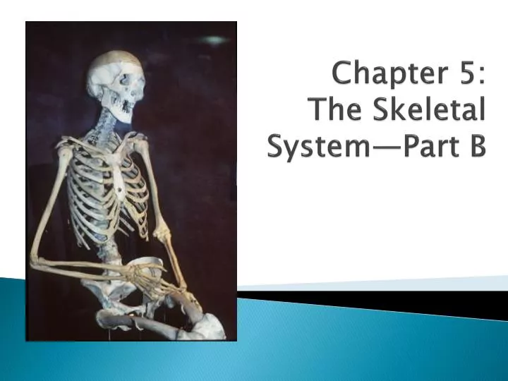
Chapter 5: The Skeletal System—Part B. Bone Fractures. Fracture - A break in a bone Types of bone fractures Closed (simple) fracture – break that does not penetrate the skin Open (compound) fracture – broken bone penetrates through the skin

E N D
Bone Fractures • Fracture - A break in a bone • Types of bone fractures • Closed (simple) fracture – break that does not penetrate the skin • Open (compound) fracture – broken bone penetrates through the skin • Bone fractures are treated by reduction (realignment of bone ends) and immobilization (cast) • 6-8 weeks healing
Common Types of Fractures Table 5.2
Repair of Bone Fractures • Hematoma (blood-filled swelling) is formed • Break is splinted by fibrocartilage to form a callus of cartilage, bone, & collagen • Fibrocartilage callus is replaced by a bony callus formed of spongy bone • Bony callus is remodeled to form a permanent patch
Hematoma Hematomaformation Stages in the Healing of a Bone Fracture Figure 5.5, step 1
Hematoma Externalcallus Internalcallus(fibroustissue andcartilage) Newbloodvessels Spongybonetrabecula Hematomaformation Fibrocartilagecallus formation Stages in the Healing of a Bone Fracture Figure 5.5, step 2
Hematoma Externalcallus Bonycallus ofspongybone Internalcallus(fibroustissue andcartilage) Newbloodvessels Spongybonetrabecula Hematomaformation Fibrocartilagecallus formation Bony callusformation Stages in the Healing of a Bone Fracture Figure 5.5, step 3
Hematoma Externalcallus Bonycallus ofspongybone Internalcallus(fibroustissue andcartilage) Newbloodvessels Healedfracture Spongybonetrabecula Bone remodeling Hematomaformation Fibrocartilagecallus formation Bony callusformation Stages in the Healing of a Bone Fracture Figure 5.5, step 4
The Axial Skeleton • Forms the longitudinal part (central line) of the body • Divided into three parts • Skull • Vertebral column • Bony thorax
The Axial Skeleton Figure 5.6
The Skull • Two sets of bones • Cranium (encloses brain) • 8 bones • Facial bones • 14 bones • Bones are joined by sutures--interlocking immovable joints • Only the mandible is attached by a freely movable joint
Cranium • 8 large flatbones • Parietal & temporal – paired • Parietal – superior & lateral • Meet midline at sagittal suture • Meet frontal bone at coronal suture • Temporal – meet parietal at squamous suture • External auditory meatus – canal to eardrum • Mastoid & styloid processes – attach neck muscles • Frontal – forehead • Occipital – posterior • Meets parietal at lambdoid suture
Bones of the Skull • Sphenoid - eye orbits & floor of cranial cavity • Optic canal – nerve to eye • Ethmoid – roof of nasal cavity & medial eye orbits • Cristagalli = cock’s comb; attaches brain • Cribriform plates – holes for olfactory nerves
The Skull Figure 5.7
Bones of the Skull Figure 5.11
Bone Markings • Sphenoid bone • Sellaturcica = Turk’s saddle • Holds pituitary gland • Foramen ovale – cranial nerves • Between temporal & occipital • Jugular foramen – junction of occipital & temporal bones • Passage of jugular vein • Temporal bone • Internal acoustic meatus – cranial nerves VII & VII • Carotid canal • For carotid artery • Occipital bone • Foramen magnum – spinal cord to brain
Human Skull, Superior View Figure 5.8
Facial Bones (14) • Lacrimal – 2; contain tear ducts • Mandible – 1; largest & strongest face bone • Maxilla – 2; fuse = upper jaw • Zygomatic – 2; cheek bones & eye sockets • Palatine – 2; • Hard palate – maxilla & palatine bones • Cleft palate if not fused • Vomer – 1; forms nasal septum • Inferior nasal conchae – 2 • Nasal - 2
Human Skull, Inferior View Figure 5.9
Paranasal Sinuses • Hollow portions of bones surrounding the nasal cavity Figure 5.10 Hoban
Paranasal Sinuses • Functions of paranasal sinuses • Lighten the skull • Give resonance and amplification to voice Figure 5.10 Hoban
Sinusitis • Recall that “-itis” means inflammation of • Sinuses are continuous with nasal passages and throat • Infections can travel up to sinuses
The Hyoid Bone • The only bone that does not articulate with another bone • Serves as a moveable base for the tongue • Aids in swallowing and speech Figure 5.12
The Fetal Skull • The fetal skull is large compared to the infant’s total body length • Adult skull – 1/8 body length • Newborn skull – ¼ body length • Fontanelles – fibrous membranes connecting the cranial bones • Allow fetal skull to be compressed during birth • Allow the brain to grow • Convert to bone within 24 months after birth Figure 5.13
The Fetal Skull Figure 5.13
Fetal Skeleton Note how large the head is compared to the rest of the body… Checkpoint: What is the advantage of a larger head area to the developing baby? More area for the brain to develop properly…