Download
1 / 64
640 likes | 774 Views
The Human Body =. HOMEOSTASIS (maintaining a constant condition). Human Circulatory System:. Also known as the cardio-vascular system Cardio refers to the heart Vascular refers to the blood vessels It is a closed system , which means that blood is confined within vessels.

E N D
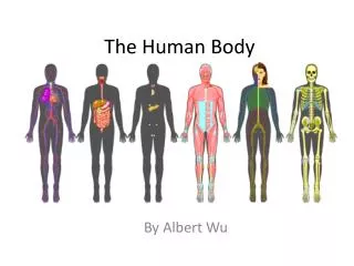
The Human Body= HOMEOSTASIS (maintaining a constant condition)
Human Circulatory System: • Also known as the cardio-vascular system • Cardio refers to the heart • Vascular refers to the blood vessels • It is a closed system, which means that blood is confined within vessels
Function of the heart: • Every cell in your body needs oxygen(O2) in order to live and function. • The purpose of the heart is to deliver the oxygen-rich blood to every cell in the body
The Atria: • Top twochambers of the heart • They have thin walls • The function of the atria are to collect blood returningto the heart from the body • The atria then send the blood directly to the ventricles through the atrio-ventricular valves
The Ventricles • Bottom two chambers • They have much thicker walls • More powerful than the atria, especially the left ventricle • The left ventricle pumps blood to the rest of the body and is therefore the strongest chamber • The right ventricle pumps blood only to the lungs to pick up oxygen (O2)
There are 3 main types of blood vessels: • Arteries • Veins • Capillaries
Arteries • Carry oxygen-rich blood AWAY from the heart to the body (exception___?) • The Aorta is the main artery leaving the heart • Arterioles (little arteries) connect larger arteries with capillaries • Small arterioles branch into collections of capillaries known as capillary beds
Veins • Carry oxygen-poor blood from capillaries back to the heart (exception __?) • Blood pressure in veins is low, so veins depend on nearby muscle contractions to move blood along • Veins are sandwiched in between skeletal muscles and whenever we move these muscles they pinch the veins and squeeze blood through
Capillaries • Thin-walled blood vessels in which gases, nutrients, wastes, and hormones are exchanged • Only one cell layer thick and microscopic in size • Blood leaving the capillary beds flow into a gradually larger series of Venules which join to form Veins. • Capillaries therefore connect arteries and veins.
Something to think about… • How many times does your heart beat in one hour? (assuming 80 bpm) • 80 x 60 = 4,800 bph • One day? • 4800 x 24 = 115,200 bpd • One week? • 115,200 x 7 = 806,400 bpw • One month? • 806,400 x 4 = 3,225,600 bpm • One year? • 3,225,600 x 12 = 38,707,200 bpy • In your lifetime? • 38,707,200 x your age (25) = 967,680,000
There are 3 different types of Circulation throughout your body: • Coronary (to the heart) • Pulmonary (to the lungs) • Systemic (to the body)
Coronary Circulation • Refers to the movement of blood through the chambers of the heart • Two small coronary arteries branch off the aorta and spread over the surface of the heart • These coronary arteries supply oxygen and nutrients to specific regions of heart muscle
Pulmonary Circulation • The movement of blood from the heartto the lungs and back to the heart again • Blood flows to the lungs to pick up oxygen and release CO2
Systemic Circulation • The movement of nutrient-rich blood to all of the tissue located throughout your body
Problems of the Circulatory System
Stroke • Results from a blockage of an artery (Carotid artery) in the head • As a result, nervous tissue in the brain dies • Can be fatal
High Blood Pressure: • Also known as hypertension • Result of reduced elasticity of artery walls which makes the heart work harder to force blood through • Can also result from a narrowing of arteries • Caused by poor diet, stress, heredity, obesity, and cigarette smoking
Heart Attack • A heart attack is when part of the heart muscle is damaged or dies because it isn't receiving oxygen. • Oxygen is carried to the heart by the arteries
Respiratory System
There are 2 types of Respiration going on in your body… • Cellular Respiration – gases entering your individual cells • Body (Systemic) Respiration – gases (O2) entering your lungs (breathing)
So, how does Oxygen get to the Lungs? • Gases are transported through the nostrils into the nasal cavity • Here the air is warmed and filtered by mucous and cilia • The air then travels down the trachea (wind pipe) into your lungs.
Cilia • The trachea & nose are lined with cilia. • Cilia sweep with a wavy motion. They move the mucus around to attract dirt and germs. • The cilia sweep the dirty mucus up toward your mouth. • Cigarette smoke slows down that motion and then stops the action of cilia, thereby allowing foreign substances to enter your lungs.
Alveoli • Bronchi divide further into bronchioleswhich are very thin and made of muscle and nerve endings • Bronchioles lead to alveoliwhich are the functional unit of the lung. • Alveoli are air sacs surrounded by capillaries and look like little bunches of grapes • Exchange of gasesoccurs at the alveoli. • There are ~ 1 Billion alveoli in the linings of your lungs!
Lungs • Eachlung contains 1 bronchi, many bronchioles, and millions of alveoli • They are highly elastic (stretchy) but have no muscle for moving air. • Instead, they respond to the action of the diaphragm muscle.
The Diaphragm • The Diaphragm is a sheet of muscle that separates the chest cavity from the abdominal cavity. • The brain and spinal cord control the movement of the diaphragm and the muscles which move the ribs. • These 2 sets of muscles control inhalation and exhalation.
Negative Feedback • The Medulla in the lower brain continuously monitors blood pH • As CO2 concentration in the blood increases, blood pH decreases (becomes more acidic). • When blood pH drops, the medulla will stimulate the diaphragm and rib muscles to contract so you can take in more O2 and release more CO2. • This process is an example of negative feedback and operates to maintain homeostasis.
Problems of the Respiratory System are… • Asthma • Lung Cancer
What is Asthma? • Asthma is a disease that causes swelling and blockage of the airways that bring air from the nose and mouth to the lungs. • We do not know what causes asthma but it tends to run in families that also have hay fever and eczema.
Asthma Triggers… • Asthma triggers are commonly related to some kind of allergic reaction, for example, to pollen or dust mites. • Other common triggers include inhalation of pollution, tobacco smoke, emotional upset, aspirin, exercise and breathingcold air.
What is Lung Cancer? • An uncontrollable growth of tumors in your lungs • Lung cancer is the number one cause of cancer deaths among men and women in the United States. • Approximately 85 to 87 % of all cases are caused by tobacco use, making lung cancer one of the most preventable cancers. • However, it is one of the most difficult to detect early, which makes treatment drastic when it is detected.
What is Digestion? • The process of breaking down large food moleculesinto smaller molecules that the body can use • There are 2 types of digestion: • Mechanical (physical) • Chemical (by enzymes)
The Mouth • Food enters the mouth and both mechanical and chemical digestionbegins. • Teeth – (mechanical digestions) - cut, tear, grind, and crush large pieces of food into smaller pieces. • Tongue – a muscle that helps move food around and helps mix food with saliva. • Saliva – (chemical digestion) – enzymes break down starches.
Chemical Digestion • Your Saliva contains enzymes which break down food. • Ex: ComplexCarbs (grains, potatoes, veggies) are broken down into simple sugars by the enzyme amylase. • Proteins are broken down into amino acids by the enzyme protease. • And fats are broken down into fatty acids and glycerol by the enzyme lipase. • Each enzyme has it’s own job that breaks down only 1 specific kind of food molecule.
The Esophagus • A short tube (~ 25 cm long) between the mouth and the stomach • Lined with mucus which helps food move easily through tube
How does Food get to the stomach? 2 WAYS!!! • Gravity – helps food move down • Peristalsis – muscle contractions help move food down the esophagus • Peristalsis can push against gravity, so that you can swallow even when you are standing on your head!
The Stomach “Churn and Burn” The stomach performing both mechanical and chemical digestion • The “burn” part refers to Chemical digestion – - Glands in the stomach release a combination of enzymes to further break down food particles. • The “churn” part refers to Mechanicaldigestion– • 3 layers of muscles contract in different directions twisting and churning the stomach contents. • As your stomach “churns” the muscles twist the stomach in three (3) different directions. • 1. Circular • 2. Diagonal • 3. Longitudinal
What does my meal look like after digestion? • By the time the stomach is done burning and churning the food for five hours, it is no longer recognizable. • The offensive line of the digestive team does such a good job on the food that it is only a white creamy, liquid known as chyme. • Chyme consists of water, starch (broken down carbs), fats and partially broken down proteins
Small Intestine (S.I.) • A narrow muscular tube about 20 feet long and 2.5 cm wide • Food moves through by peristalsis (remember this from the esophagus???) • Chemical digestion of starches, proteins, and fats is completed in the S.I by digestive juices secreted from the liver, pancreas, and the SI itself
Villi • Absorption of the broken down food takes place in the wall of the SI • The inner lining is folded and has millions of tiny fingerlike projections called VILLI • These villi help food pass into the bloodstream
The Large Intestine (LI) is made up of 3 parts: • The Cecum • The Colon • The Rectum
The Colon • Like the small intestine, the colon section of the LI is twisted around the abdomen . • The food in the colon is now considered to be waste, and is waiting to be kicked out. • The function of the colon is to absorb water to maintain the consistency of the feces.
The Rectum • The first part of the rectum is called the rectal cavity, its other parts are the sphincter and the anus. • Waste or feces that are expelled from the large intestine have no nutritional value. • The body has already taken everything out of the food that it can use. • And is now waiting to be expelled from the body completely.
Problems of the Digestive System
Ulcer • When stomach acids eat away at the lining of the stomach and create a hole or sore • Ulcers are generally caused by an imbalance between pepsin (enzyme) and stomach acid • Can be caused by an infection of the bacteria Helicobacter Pylori which changes the stomachs mucus layer causing an ulcer.