Download
1 / 16
500 likes | 1.67k Views
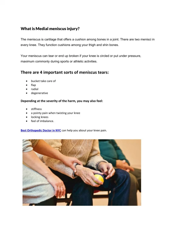
Medial meniscus injury. Function of menisci. Acts as the shock absorber Helps the knee in locking mechanism Assists and controls gliding and rolling motion of the knee Contributes towards the stability of the knee joint Weight transmission. Medial meniscus injury .

E N D
Function of menisci • Acts as the shock absorber • Helps the knee in locking mechanism • Assists and controls gliding and rolling motion of the knee • Contributes towards the stability of the knee joint • Weight transmission
Medial meniscus injury • Medial meniscus is more commonly injuried than the lateral and is usually associated with other ligament injuries of the knee. • Lateral meniscus is less common : - because it is smaller in diameter , thicker in periphery , wide , more mobile and attached to both cruciate ligament.
Mechanism of injury • Rotational force when a flexed knee extends • In young : it can occur only when weight is being taken , knee is flexed and there is a twisting strain. Young active athletes • In middle life : fibrosis has decreased the mobility of meniscus and hence tear occurs with less force
Features medial meniscus Shape - semicircular Anterior horn - attached to tibial intercondylar eminence in front ACL Posterior horn – intercondylar area in front of PCL and behind posterior horn of lateral meniscuc Mobility - less mobile Lateral meniscus - circular - attached to tibial intercondylar eminence lateral to ACL To the intercondylar eminence more mobile
classification based on their tear patterns : - Diagram of meniscal tear patterns: (A) Vertical or longitudinal (Bucket-handle), (B) Flap or Oblique, (C) Radial or Transverse, (D) Horizontal, (E) Complex degenerative
Most common symptoms • Knee pain • Swelling of the knee • Tenderness when pressing on the meniscus • Popping or clicking within the knee • Limited motion of the knee joint
Signs • Locking +ve • McMurray`s test +ve • Apley`s test +ve • Duck waddle test +ve • Steinmann`s sign +ve • Quadriceps atrophy +ve • Medial joint line tenderness +ve
Figure 5. The McMurray test Is performed by flexing the patient's hip and knee and palpating for a pop or click along the joint line as the tibia is internally and externally rotated
Figure : - The Apley test Apley`s distraction test Apley`s compression test
Figure 4. The Steinman test produces joint line pain when the tibia is rotated internally and externally while the knee is flexed over the examination table