Download
1 / 23
310 likes | 553 Views
Thyroid Gland Parathyroid Trachea Esophagus By Prof . Saeed Abuel Makarem. VISCERA OF THE NECK. The deep cervical fascia of the neck is divided into 3 layers: 1- Investing layer. 2- Pretracheal layer. 3- Prevertebral layer. Thyroid gland. Endocrine gland.

E N D
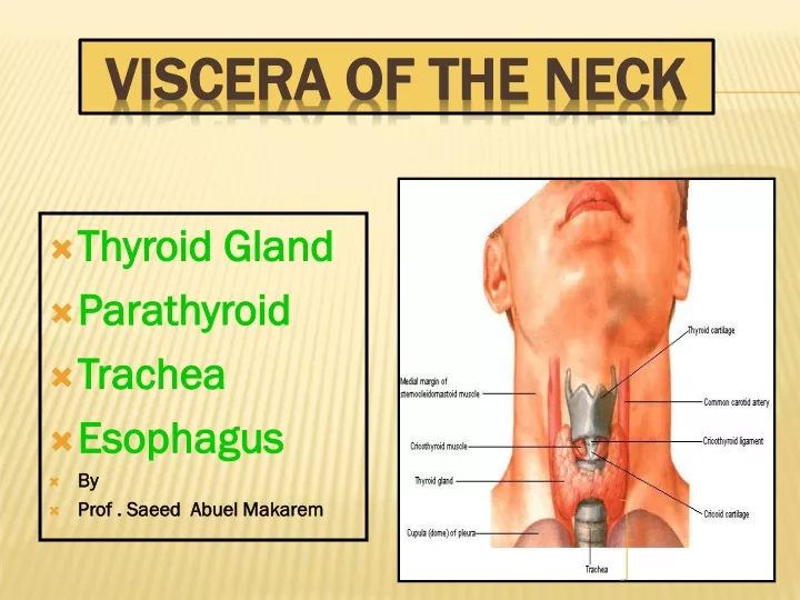
Thyroid Gland Parathyroid Trachea Esophagus By Prof . Saeed Abuel Makarem VISCERA OF THE NECK
The deep cervical fascia of the neck is divided into 3 layers: • 1- Investing layer. • 2- Pretracheal layer. • 3- Prevertebral layer.
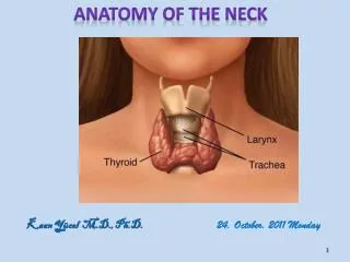
Thyroid gland • Endocrine gland. • Consists of right & left lobes. • The 2 lobes are connected to each other by a narrow isthmus, which overlies the 2nd 3rd & 4th rings of the trache. • It is surrounded by a sheath derived from the pretracheal layer of cervical fascia.
Each lobe is pear shaped, with its apex reaches up to the oblique line of the thyroid cartilage. • Its base lies at the level of 4th or 5th tracheal rings. • Inside the pretracheal facial capsule, there is another capsule.
Each lobe is pear shaped, with its apex directed upward as for as the oblique line of the thyroid cartilage; its base is at the 4th or 5th tracheal ring. The isthmus extends across the midline in front of the 2nd 3rd & 4th tracheal rings.
A small pyramidal lobe is often present which projects from the upper border of the isthmus usually to left of middle line. • Pyramidal lobe is connected to hyoid bone by a fibrous or muscular band called levator glandulaethyroideae. • This represents the fibrosed & obliterated thyroglossal duct.
Anterolaterally: • Sternothyroid. • Superior belly of Omohyoid • Sternohyoid. • Sternomastoid. • Posterolaterally: • Carotid sheath & its contents. • Medially: • Above: • Larynx & pharynx . • Below: • Trachea & esophagus. • Recurrent laryngeal nerve in between. • Cricothyroid muscle & external laryngeal nerve.
Relation of the isthmus • Anteriorly: • sternothyroid, sternohyoid, • anterior jugular vein, fascia & skin. • Posteriorly: • 2nd,3rd,&4th tracheal rings. • Terminal branches of the 2 superior thyroid arteries which anastomosis along the upper border.
The rounded posteriorborder is related to the superior & inferior Parathyroid glands. • It is also related to the anastomosis between superior & inferior thyroid arteries.
: Superior thyroid artery • From external carotid artery • It descends to the upper pole of the lobe, with the external laryngeal nerve. • It runs along the upper border of the isthmus to anastomosis with its fellow. Thyroideaima artery • If present, it arises from aortic arch or from brachiocephalic artery. • It ascends in front of trachea to reach isthmus.
Inferior thyroid artery • From thyrocervical trunk of 1st part of subclavianartery, ascends behind the gland to the level of cricoid cartilage. • Then it turns medially behind the carotid sheath. • The it reaches the posterior border of the gland & descends downwards. • The recurrent laryngeal nerve crosses either in front or behind the artery.
Veins of Thyroid Gland 1- Superior thyroid vein internal jugular 2- Middle thyroid vein internal jugular 3- Inferior thyroid vein left brachiocephalic Lymph Of the Thyroid Gland: Deep cervical & paratracheal
Goiter GOITER A non-neoplastic & non-inflammatory enlargement of the thyroid gland.
Ectopic Thyroid tissue • The thyroid glands develops high up close to foramen cecum of the developing tongue. • Then it descends along the thyroglossal duct to reach its final position by the 7th week. • Descent of the thyroid could be arrested at any point, or extends down to thorax.
Parathyroid glands • Four small ovoid bodies, about 6mm. Long. • They lie within the facial capsule of the gland. • 2 superior parathyroid has a constant position at the middle of posterior border of the gland. • 2 inferior parathyroid usually at the level of the inferior pole. • They lie within the thyroid tissue or sometimes outside the facial capsule.
PARATHYROID GLAND • They are supplied by superior & inferior thyroid arteries. • Their veins are drained to superior, middle and inferior thyroid veins. • Lymph nodes: • Deep cervical & paratracheal lymph nodes. • Nerve supply: • Superior & middle cervical sympathetic ganglia.
TRACHEA • 10 to 15 cm long mobile tube. • Formed of cartilage & membrane. • Its diameter is about 2 cm in adult male. • It begins at lower border of cricoid cartilage (C 6). • It descends in the midline of the neck. • It ends at the level of the disc between T4 & T5. • .
The trachea has a fibro -elastic wall which is supported by series of U-shaped bars of hyaline cartilage that keep the lumen patent.
The posterior free ends of the cartilage are connected by smooth muscle, called the trachealismuscle.
RELATION • Anteriorly: • Skin, • Superficial fascia, investing cervical fascia • isthmus of thyroid gland, • Inferior thyroid veins • jugular arch, • thyroidea ima artery, • left brachiocephalic vein, • sternothyroid & sternohyoid
TRACHEA • Posteriorly • esophagus, • recurrent laryngeal nerves • (in between trachea and esophagus) • vertebral column. • Laterally: • Lobes of the thyroid gland & • carotid sheath. • Blood supply: • Inferior thyroid artery. • Nerve : • vagi, sympathetic trunk & recurrent laryngeal • Lymph: Pretracheal & Para tracheal lymph nodes.
ESOPHAGUS • Muscular tube 25 cm or 10 inches long. • Extends from pharynx to stomach. • It begins at lower border of cricoid cartilage (C 6). • It begins in midline, but inclines to the left. • It descends in superior then posterior mediastinum of thorax. • Anteriorly • trachea and recurrent laryngeal nerves.
ESOPHAGUS • Posteriorly • Prevertebral layer of cervical fascia, • longus coli muscle & • vertebral column. • Laterally: • Lobe of thyroid gland, • carotid sheath. • Thoracic duct on left side. • Blood: • Inferior thyroid artery. • Lymph: • deep cervical lymph nodes. • Nerves: Recurrent laryngeal and sympathetic trunk.