Download
1 / 11
110 likes | 359 Views
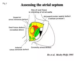
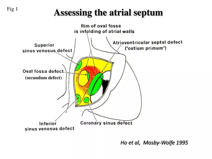
Fig 1. Assessing the atrial septum. (secundum defect). Ho et al, Mosby-Wolfe 1995. Fig 2. Secundum ASD. Apical four chamber view demonstrating a large secundum atrial septal defect. Subcostal four chamber view of same patient confirming a left to right shunt at atrial level. Fig 3.

E N D
Fig 1 Assessing the atrial septum (secundum defect) Ho et al, Mosby-Wolfe 1995
Fig 2 Secundum ASD Apical four chamber view demonstrating a large secundum atrial septal defect Subcostal four chamber view of same patient confirming a left to right shunt at atrial level
Fig 3 Secundum ASD 3D image of secundum ASD and image during transcatheter occlusion
Fig 4 TOE secundum ASD LA RA Transesophageal longitudinal view of the atria demonstrating an ostium secundum defect. Colour flow image from the same view demonstrating left-to-right shunting across the ASD
Fig 5 Primum ASD LV RV RA LA Apical four chamber view demonstrating a primum atrial septal defect Colour Doppler flow image from same view illustrating left-to-right shunt across the primum atrial septal defect
Fig 6 Parasternal short axis view demonstrating tri-leaflet left atrioventricular valve in a patient with a primum atrium septal defect
Fig 7 Primum ASD Subcostal four chamber view demonstrate an large primum atrial septal defect which is located at the low part of normal atrial septum and the junction with atrioventricular valve. LA RA LV RV
Fig 8 Primum ASD repaired LV LA Apical four chamber view of patient with a primum ASD repair. Note the thickened left AV valve leaflet and markedly dilated left atrium Colour Doppler flow mapping of the same patient demonstrating severe left AV valve regurgitation.
Fig 9 SVC type ASD • Apical four chamber view showing a sinus venous defect of the superior vena caval type
Fig 10 SVC type ASD (TOE) LA RA SVC Colour flow image from the same view demonstrating left-to-right shunting across the ASD Transesophageal longitudinal view of the atria demonstrating a sinus venousus defect of the superior vena caval type.
Fig 11 Sinus venousis ASD (SVC type) Transesophageal longitudinal view of the atria demonstrating a sinus venosus defect of the superior vena caval type.